| 包装: | 100mg |
| 市场价: | 446元 |
Cell experiment: | The cells are resuspended and stained for 30 min in 1.0 mL of 0.01% pyronin Y in sodiumacetate buffer (1.0 M, pH 4.7). For this purpose 1.0 g of pyronin Y is dissolved in 100 mL distilled water and this solution is purified by chloroform extraction (three fractions of 100 mL). The purified pyronin Y solution is diluted with 1.0 M sodiumacetate buffer pH 4.7 to the appropriate concentration and filtered through paper filters before use. Stained cell samples are studied by fluorescence microscopy (excitation filters SP 560 and LP 515, chromatic beam splitter at 580 nm, barrier filter LP 580) or by flow cytometry[2]. |
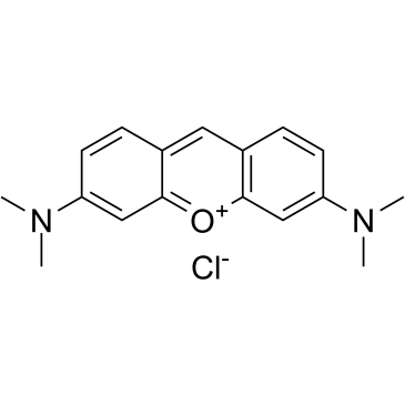
| 产品描述 | Pyronin Y is an intercalating cationic dye that shows specificity towards RNA. Pyronin Y forms fluorescent complexes with double-stranded nucleic acids, especially RNA, enabling semi-quantitative analysis of cellular RNA in flow cytometry, to estimate the RNA content per cell in formalin fixed EL4 leukosis tumor cells, enzyme dispersed R3327-G rat prostatic adenocarcinoma cells, mouse spleen cells stimulated with concanavalin A, and human peripheral blood lymphocytes stimulated with phytohemagglutinin[1]. A fluorescent staining procedure based on pyronin Y is described. The technique has been used to stain RNA in human reticulocytes for subsequent flow analysis and sorting[2]. In viable cells this dye also accumulates in mitochondria. At a concentration of 1.7 to 3.3 μM, pyronin Y is localized almost exclusively in mitochondria of cultured cells, similar to another mitochondria! probe, rhodamine 123. At that concentration PY is not toxic but suppressed cell growth, arresting cells[3]. Pyronin Y has long been used, in combination with other dyes such as Methyl Green, as a differential stain for nucleic acids in paraffin tissue sections. In sections stained with Methyl Green-pyronin Y, red blood cells, elastic fibre of blood vessels, zymogen granules of pancreatic acinar cells, surface membrane of heptocytes and kidney tubular cells showed strikingly strong green and/or red fluorescence, while the nuclei of cells appeared non-fluorescent[4]. [1]. Pollack A, et al. Flow cytometric analysis of RNA content in different cell populations using pyronin Yand methyl green. Cytometry. 1982 Jul;3(1):28-35. [2]. Tanke HJ, et al. Flow cytometry of human reticulocytes based on RNA fluorescence. Cytometry. 1981 Mar;1(5):313-20. [3]. Darzynkiewicz Z, et al. Cytostatic and cytotoxic properties of pyronin Y: relation to mitochondrial localization of the dye and its interaction with RNA. Cancer Res. 1986 Nov;46(11):5760-6. [4]. Li B, et al. Pyronin Y as a fluorescent stain for paraffin sections. Histochem J. 2002 Jun-Jul;34(6-7):299-303. |
 m.cnreagent.com