| CAS NO: | 2009273-71-4 |
| 规格: | ≥98% |
| 包装 | 价格(元) |
| 5mg | 电议 |
| 10mg | 电议 |
| 25mg | 电议 |
| 50mg | 电议 |
| 100mg | 电议 |
| 250mg | 电议 |
| Molecular Weight (MW) | 348.406 |
|---|---|
| Formula | C20H20N4O2 |
| CAS No. | 2009273-71-4 ;2009273-60-1 |
| Storage | -20℃ for 3 years in powder form |
| -80℃ for 2 years in solvent | |
| Solubility (In vitro) | DMSO: ≥ 310 mg/mL |
| Water: <1 mg/mL | |
| Ethanol: 1 mg/mL | |
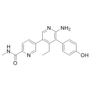
| SMILES | O=C(C1=CC=C(C2=C(CC)C(C3=CC=C(O)C=C3)=C(N)N=C2)C=N1)NC |
| Synonyms | GNE-6776; GNE6776; GNE 6776 |
| In Vitro | In vitro activity: High-throughput screen and counterscreen assays. USP7 UbA10 time-resolved fluorescence energy transfer (TR-FRET) activity assay. Potential inhibitors of USP7 were identified in a TR-FRET-based enzyme activity assay with UbA10 as substrate. UbA10 is a fragment of the naturally occurring ubiquitin precursor Ub52; it retains the ten-amino-acid segment of Ub52 that extends beyond the ubiquitin C terminus. The primary screening assay is a novel TR-FRET-based activity assay that measures cleavage by full-length USP7 of a doubly tagged peptide substrate. The peptide is tagged with glutathione S-transferase (GST) on the N terminus and with eight histidine residues on the C terminus (GST–UbA10-His). The tags are detected by anti-GSH-d2 (TR-FRET acceptor) and anti-His-europium (TR-FRET donor), respectively. Cleavage of this substrate by USP7 at the ubiquitin C terminus results in separation of these two tags and loss of TR-FRET signal. Compounds were dispensed into 1,536-well black plates (MaKO, Aurora Microplates) followed by 2 μ l full-length recombinant USP7 in assay buffer (50 mM HEPES pH 7.5, 0.1% Prionex (Pentapharm), 0.01% Triton X-100, and 10 mM dithiothreitol (DTT)). After a 10-min incubation, the reaction was started by the addition of 2 μ l GST–UbA10-His substrate in assay buffer. After 75 min of reaction, 2 μ l of a detection antibody reagent, containing anti-His-europium (Life Technologies) and anti-GST-d2 (CISbio) in assay buffer were added. After 60 min, the fluorescence at 618 nm and 671 nm with excitation at 340 nm was read on a ViewLux reader (PerkinElmer). Cleavage of the doubly tagged substrate resulted in loss of TR-FRET signal, whereas inhibition of USP7 by compound restored the signal. The TR-FRET ratio was calculated as fluorescence at 671 nm divided by fluorescence at 618 nm. TR-FRET ratios were normalized to controls to determine the percentage inhibition for the single concentration screen. For confirmation in concentration–response mode (ten points with n = 2), percentage inhibition was plotted against compound concentration, and the data were fitted to a four-parameter curve with Screener Assay Analyzer (Genedata) to determine IC50 values. The assay buffer was optimized to maintain enzyme stability and to maximize assay signal to background: Triton X-100 was included to prevent non-specific adsorption of the enzyme and/or substrate to the assay plate and/or to compound aggregates, Prionex carrier protein was included to help stabilize the enzyme and to prevent non-specific adsorption to container and tubing surfaces as well as to minimize non-specific inhibition by library compounds, and DTT served to maintain good USP7 activity and minimize the impact of inhibitors that act through redox cycling. Reagent concentrations were optimized for good assay performance at approximately 50% substrate conversion in the signal decrease assay with a key aim of minimizing the required concentration of USP7. The GST–UbA10-His concentration was adjusted to maximize assay signal, anti-GST-d2 concentration was adjusted approximately in parallel with that of the substrate, and anti-His-europium was used at a concentration that represented a minimum that was compatible with robust detection on the ViewLux plate reader. Time-course evaluations were conducted to confirm that the enzyme concentration (10 nM) and reaction time (75 min) were in the linear range of enzyme activity. Additionally, extended time courses for the detection reaction were used to demonstrate that the 60-min incubation was sufficient to reach equilibrium. Under the final assay conditions, reagent stability studies indicated greater than 20 h of acceptable performance. Over the course of the screen, Z′ values averaged 0.76. Kinase Assay: USP7 di-ubiquitin FRET activity assay. Potential USP7 inhibitors were confirmed in an orthogonal activity assay with an internally quenched K63-linked di-ubiquitin substrate (U-310, Boston Biochem). Conditions were similar to those used for the UbA10 TR-FRET activity assay. Compounds dispensed into 1,536-well plates were preincubated with full-length recombinant USP7 in assay buffer for 10 min, and the reaction was started by the addition of 2 μ l of the di-ubiquitin substrate. During the 60-min incubation, cleavage of the substrate by USP7 resulted in the release of quenching of the TAMRA tag by the QXl tag and thus an increase in fluorescence. The fluorescence intensity was read with excitation at 540 nm and emission at 585 nm. Concentration–response assay methods and data analyses were conducted as for the UbA10 TR-FRET assay. USP7 Ubiquitin/Rho110 activity assay. Potential USP7 inhibitors were also confirmed in an orthogonal activity assay with ubiquitin/rhodamine-110 as substrate. Compounds were preincubated for 10 min with 2 μ l full-length recombinant USP7 in assay buffer, and the reaction was started by the addition of 2 μl of ubiquitin/rhodamine-110 (U-555, Boston Biochem). After a 60-min reaction in which USP7 cleaved the rhodamine-110 from the ubiquitin and thus increases fluorescence, the fluorescence intensity was read with excitation at 485 nm and emission at 535 nm. General assay conditions and data analyses were as described Cell Assay: EOL-1 cells are seeded into 384-well plates 24 h before compound addition. Cells are then incubated with compound (e.g., GNE-6776; 0.003, 0.009, 0.027, 0.082, 0.25, 0.74, 2.22, 6.67, and 20 μM) for 72 h or 120 h before assaying viability. Assays are performed in biological triplicate. Cells are incubated (37°C, 5% CO2) in RPMI-1640, 2.5% FBS (72 h assay) or 5% FBS (120 h assay), and 2 mM glutamine throughout the assay. The reported IC50 and mean viability metrics are as follows: IC50 is the dose at which the estimated inhibition is 50% relative to untreated wells (that is, absolute IC50). The mean viability is calculated |
|---|---|
| In Vivo | GNE-6776 promotes on-target pathway modulation in human xenografts. Although efficacious exposure is only transiently achieved, GNE-6776 causes modest, although significant, EOL-1 xenograft growth delay |
| Animal model | Female C.B-17 SCID mice |
| Formulation & Dosage | 200 mg/kg (body weight) by oral gavage |
| References | Nature. 2017 Oct 26;550(7677):534-538. |
 m.cnreagent.com