Ripasudil 是一种ROCK特异性抑制剂,能够抑制ROCK1和ROCK2的活性,IC50值分别为 51 和 19 nM。
产品描述
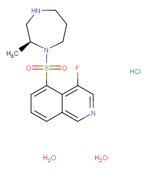
Ripasudil (K-115) hydrochloride dihydrate is a specific ROCK inhibitor (IC50s: 51/19 nM for ROCK1/ROCK2).
体外活性
Ripasudil shows less potent inhibitory activities against CaMKIIα, PKACα, and PKC (IC50s: 370 nM, 2.1 μM and 27 μM) [1]. Ripasudil (1, 10 μM) induces cytoskeletal changes, including retraction and cell rounding and reduced actin bundles of cultured trabecular meshwork (TM) cells. Ripasudil (5 μM) significantly reduces transendothelial electrical resistance (TEER) and increases FITC-dextran permeability in Schlemm's canal endothelial (SCE) cell monolayers [2].
体内活性
Ripasudil reduces intraocular pressure in a concentration-dependent manner at concentrations between 0.1% and 0.4% in monkey eyes and 0.0625% to 0.5% in rabbit eyes, respectively [1]. Ripasudil (1 mg/kg, p.o. daily) shows a neuroprotective effect on retinal ganglion cells (RGCs) after nerve crush (NC). Ripasudil also inhibits the oxidative stress induced by axonal injury in mice. Ripasudil suppresses the time-dependent production of ROS in RGCs after NC injury [3].
激酶实验
ROCK 1 (0.75 ng/mL) and ROCK 2 (0.5 ng/mL) are incubated with various concentrations of Ripasudil, Y-27632, or HA-1077 at 25°C for 90 min in 50 mM Tris-HCl buffer (pH 7.5) containing 100 mM KCl, 10 mM MgCl2, 0.1 mM EGTA, 30 mM Long S6 Kinase Substrate peptide, and 1 mM ATP in a total volume of 40 mL. PKACa, PKC, and CaMKIIa are also incubated with various concentrations of Ripasudil, Y-27632, or HA-1077. PKACa (0.0625 ng/mL) is incubated at 25°C for 30 min in 40 mM Tris-HCl buffer (pH 7.5) containing 20 mM MgCl2, 1 mg/ mL BSA, 5 mM Kemptide peptide substrate, and 1 mM ATP in a total volume of 40 mL. PKC (0.025 ng/mL) is incubated at 25°C for 80 min in 20 mM Tris-HCl buffer (pH 7.5) containing 20 mM MgCl2, 0.4 mM CaCl2, 0.1 mg/mL BSA, 0.25 mM EGTA, 25 ng/mL phosphatidylserine, 2.5 ng/mL diacylglycerol, 0.0075% Triton-X-100, 25 mM DTT, 10 mM Neurogranin (28-43) peptide substrate, and 1 mM ATP in a total volume of 40 mL. CaMKIIa (0.025 ng/mL) is incubated at 25°C for 90 min in 50 mM Tris-HCl buffer (pH 7.5) containing 10 mM MgCl2, 2 mM CaCl2, 0.04 mg/mL BSA, 16 mg/mL purified calmodulin from bovine testis, 500 mM DTT, 50 mM Autocamitide 2, and 1 mM ATP in a total volume of 40 mL. After incubation, 40 mL of KinaseGlo Luminescent Kinase Assay solution is added, and allowed to remain at 25°C for 10 min, and Relative Light Units (RLU) are measured using a luminometer. The RLU without test compound is set as 100% (Control value), and that without enzyme and compound is set as 0% (Normal value). The reaction rate (% of control) is then calculated from the RLU with addition of each concentration of test compounds, and the 50% inhibitory concentrations (IC50) are determined by logistic regression analysis using SAS [1].
细胞实验
Trabecular meshwork (TM) cells are plated on 6 well plates at a density of 1?×?10^4 cells per well in DMEM containing 10% FBS. Following overnight culture, when cells have reached semiconfluence, 1 or 10?μM of Ripasudil, 10?μM of Y-27632, or 10?μM of fasudil are added to culture wells. PBS is used as a control vehicle. After 60?min, drug solutions are removed and replaced with DMEM containing 10% FBS. Cells are observed by phase-contrast microscopy and photographed 60?min after drug application and 2?h after drug removal. For immunohistochemistry, TM cells are plated on gelatin-coated 8 well chamber slides at a density of 1?×?10^4 cells per well in DMEM containing 10% FBS. After overnight culture, when cells reach semiconfluence, cells are incubated in Ripasudil at 1 or 10?μM, Y-27632 at 10?μM, or fasudil at 10?μM for 60?min. PBS is used as a control vehicle. Drug solutions are removed and replaced with DMEM containing 10% FBS after 2?h. Cells are fixed with 4% paraformaldehyde in PBS for 15?min then washed with cytoskeletal buffer (10?mM MES, 150?mM NaCl, 5?mM EGTA, 5?mM MgCl2, 5?mM glucose, pH 6.1) and serum buffer (10% FBS in PBS). Cells are permeabilized with 0.5% Triton X-100 in PBS for 12?min at room temperature and blocked with serum buffer for at least 2?h at 4°C. Filamentous actin (F-actin) is labeled with 0.05?mg/mL Phalloidin-TRITC for 1?h at room temperature. After washing with PBS, cells are mounted with a commercial mounting medium containing DAPI and observed using a fluorescence microscope. The exposure to take images for F-actin and DAPI are 0.1 and 0.05?sec, respectively [2].
动物实验
In the rabbit experiments, 50 mL of vehicle or Ripasudil at concentrations of 0.0625%, 0.125%, 0.25, or 0.5% is instilled into one eye. Intraocular pressure (IOP) is measured in both eyes before and 0.5, 1, 2, 3, 4, and 5 h after instillation. The contralateral eye is not treated. Animals are administered all concentrations of Ripasudil assigned using the Latin square method with intervals of at least 2 d. In the monkey experiments, 20 mL of Ripasudil at concentrations of 0.1%, 0.2%, or 0.4%, and latanoprost at a concentration of 0.005% are instilled into one eye. IOP is measured in both eyes before and 1, 2, 4, 6, and 8 h after instillation. The contralateral eye is not treated. Animals are arranged to receive all formulations with intervals of at least 1 week using the Latin square method. The IOPs are compared with the results for the instillation side at pre-dose and at each time point after the instillation of Ripasudil and are compared with both eyes at each time point.
Cas No.
887375-67-9
分子式
C15H21ClFN3O3S
分子量
377.86
别名
K-115;Ripasudil hydrochloride dihydrate;瑞舒地尔盐酸二水合物
储存和溶解度
H2O:45 mg/mL (113.67 mM)
Powder: -20°C for 3 years
In solvent: -80°C for 2 years
 m.cnreagent.com