| CAS NO: | 940929-33-9 |
| 规格: | ≥98% |
| 包装 | 价格(元) |
| 5mg | 电议 |
| 10mg | 电议 |
| 25mg | 电议 |
| 50mg | 电议 |
| 100mg | 电议 |
| 250mg | 电议 |
| 500mg | 电议 |
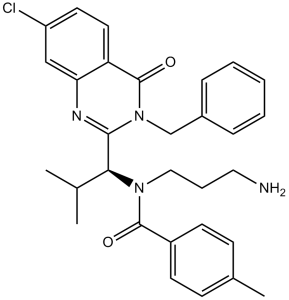
| Molecular Weight (MW) | 553.52 |
|---|---|
| Formula | C31H33N2O3.HCl |
| CAS No. | 940929-33-9 (HCl) |
| Storage | -20℃ for 3 years in powder form |
| -80℃ for 2 years in solvent | |
| Solubility (In vitro) | DMSO: 111 mg/mL (200.5 mM) |
| Water: 22 mg/mL (39.7 mM) | |
| Ethanol: 111 mg/mL (200.5 mM) | |
| Solubility (In vivo) | Saline pH5.0: 30 mg/mL |
| Synonyms | SB-743921; SB743921; SB 743921; SB-921; SB 921; SB921; GSK 921; GSK921; GSK-921; GSK921; GSK-743921; GSK 743921; GSK743921 |
| In Vitro | In vitro activity: The Ki of SB 743921 for human and mouse KSP is 0.1 nM and 0.12 nM, respectively, while the Ki of SB 743921 for other kinesins including MKLP1, Kin2 is more than 70 μM. SB 743921 blocks assembly of a functional mitotic spindle, thereby causing cell cycle arrest in mitosis and subsequent cell death. SB-743921 has improved potency over ispinesib in both biochemical and cellular assays. Kinase Assay: The motor domains of KSP (amino acids 1–360) is expressed as in Escherichia coli BL21(DE3) as COOH-terminal 6-his fusion proteins. Bacterial pellets are lysed in a microfluidizer with a lysis buffer [50 mM Tris-HCl; 50 mM KCl, 10 mM imidazole, 2 mM MgCl2, 8 mM β-mercaptoethanol, 0.1 mM ATP (pH 7.4)], and proteins are purified using Ni-NTA agarose affinity chromatography, with an elution buffer consisting of 50 mM PIPES, 10% sucrose, 300 mM imidazole, 50 mM KCl, 2 mM MgCl2, mM β-mercaptoethanol, and 0.1 mM ATP (pH 6.8). Steady-state measurements of ATPase activity are performed with a pyruvate kinase–lactate dehydrogenase detection system that coupled the appearance of ADP with oxidation of NADH. Absorbance changes are monitored at 340 nm. All biochemical experiments are performed in PEM25 buffer [25 mM Pipes/KOH (pH 6.8), 2 mM MgCl2, 1 mM EGTA] supplemented with 10 μM SB 743921 for experiments involving microtubules. Rates of ADP release are measured in a stopped-flow apparatus; the decrease in fluorescence of MANT-ATP is monitored. Rates of Pi release are measured in a stopped-flow apparatus, using bacterial phosphate binding protein modified with 7-diethylamino-3-((((2 maleimidyl)ethyl)amino)carbonyl)coumarin (MDCC) dye. Ki estimates of KSP inhibitors are extracted from the dose–response curves, with explicit correction for enzyme concentration. Tubulin polymerization by measuring changes in absorbance at 340 nm is monitored. The assay is performed in 100-μL volumes in 96-well half-area microtiter plates, using a microplate reader with the incubation temperature set at 37 °C. Cell Assay: All cells including HeLa cells are cultured in 10% FCS in RPMI 1640 in 5% CO2. We assessed 48-hour growth inhibition by serial dilution of SB 743921 relative to DMSO-treated cells in 96-well microtiter plates, using 3-(4,5-dimethylthiazol-2-yl)-5-(3-carboxymethoxyphenyl)-2-(4-sulfophenyl)-2H-tetrazolium. Cell growth is represented as the ratio of absorbance of treated cells to DMSO control, plotted by concentration and fitted to a four-parameter curve. Concentrations at which cellular growth is inhibited by 50% are extrapolated from the curve fit. The DNA content of HeLa cells cultured in the presence or absence of 1 μM SB 743921 for 24 hours is assessed by propidium iodide staining and flow cytometry. Immunofluorescence images are collected of HeLa cells treated for 24 hours with 1 μM SB 743921, fixed with 2% formaldehyde, permeabilized, and stained with DM1-α, anti-γ-tubulin, and 1 μg/mL 4′,6-diamidino-2-phenylindole, and with Alexa 488 secondary goat antirabbit IgG and Rhodamine-X goat antimouse IgG. Images are collected with a DeltaVision Restoration Microscopy System at a magnification of ×600. Z stacks (0.2 μm) are collected, and out of focus information is removed by constrained iterative deconvolution. Z stacks are then compressed into to a single image plane. |
|---|---|
| In Vivo | SB-743921 is greater efficacy in vivo against P388 leukemia. SB-743921 has significant efficacy in a broad spectrum of tumor models differing from that of taxanes. SB-743921 is shown to have activity against advanced human tumor xenografts Colo205 (complete regressions), MCF-7, SK-MES, H69, OVCAR-3 (complete and partial regressions), and HT-29, MX-1, MDA-MB-231, A2780 (tumor growth delay). SB-743921 doesn't cause the neuropathy often associated with the tubulin agents. |
| Animal model | Female BDF1 mice with P388 lymphocytic leukemia cells |
| Formulation & Dosage | Dissolved in 2% dimethylacetamide + 2% Cremophor EL + 96% acidified water [pH 5.0]; 10 7.5 mg/kg- 30 mg/kg; i.p. injection |
| References | Cancer Res. 2004 May 1;64(9):3276-80. |
 m.cnreagent.com