| CAS NO: | 667463-85-6 |
| 规格: | ≥98% |
| 包装 | 价格(元) |
| 5mg | 电议 |
| 10mg | 电议 |
| 25mg | 电议 |
| 50mg | 电议 |
| 100mg | 电议 |
| 250mg | 电议 |
| 500mg | 电议 |
Molecular Weight (MW) | 398.21 |
Formula | CC18H12BrN3O3 |
CAS No. | 667463-85-6 |
Storage | -20℃ for 3 years in powder form |
-80℃ for 2 years in solvent | |
Solubility (In vitro) | DMSO: 19 mg/mL (47.7 mM) |
Water: <1 mg/mL | |
Ethanol: <1 mg/mL | |
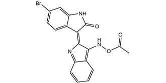
SMILES | O=C1NC2=C(C=CC(Br)=C2)/C1=C3NC4=C(C=CC=C4)C/3=N\OC(C)=O |
Synonyms | BIA; GSK-3 Inhibitor X;6-Bromoindirubin-3′-acetoxime; BIO-acetoxime; (2′Z,3′E)-6-Bromoindirubin-3′-acetoxime, 6-Bromoindirubin acetoxime, 6-Bromoindirubin-3′-acetoxime, |
In Vitro | In vitro activity: In human oral epithelial cells, BIO-acetoxime suppresses viral gene expression and protects oral epithelial cells from HSV-1 infection. In SY5Y-MYCN cells, BIO-acetoxime strongly reduces c-MYC expression and p-SMAD3 levels. BIO-acetoxime also decreases cell viability of KCN, KCNR, SY5Y, Kelly, and IMR32 cells by mediating apoptosis. In HEK 293T cells, BIO-acetoxime is also found to reduce antiviral innate immunity downstream of IRF3 activation by inhibition of GSK3α/β activities.
Kinase Assay: BIO-acetoxime (BIA) is a potent and selective GSK-3 inhibitor, with IC50s of both 10 nM for GSK-3α/β. BIO-acetoxime has anticonvulsant and anti-infection activity.
Cell Assay: OC3 cells are seeded in 3.5- and 6-cm diameter dishes for western blot analysis and RNA isolation, respectively. Cells are mock treated or exposed to HSV-1 at a multiplicity of infection (m.o.i.) of 5 unless otherwise specified. The glycogen synthase kinase-3 (GSK-3) inhibitor BIO-acetoxime (BIA), is first dissolved in DMSO to make a stock solution. Medium containing DMSO serves as a solvent control. Cells are pretreated with medium only, medium containing 0.1 % DMSO or BIO-acetoxime (BIA) for 45 min prior to mock or HSV-1 infection. BIO-acetoxime (BIA) is also present throughout the infection period. To examine the direct effect on viruses, HSV-1 is treated directly with medium containing 5 μM BIO-acetoxime (BIA) for 1 h at room temperature. Cell morphology is observed with an inverted microscope. |
In Vivo | BIO-acetoxime (BIA) shows anticonvulsant effects in the focal pilocarpine rat model at 0.5 mg/kg, and in 6-Hz fully kindled FVB/N mice at 0.5, 2.5, and 5 mg/kg via i.p. administration. |
Animal model | Mice: Male FVB/N mice are used to test the anticonvulsant effects of BIO-acetoxime in fully kindled mice. To achieve the fully kindled state, mice are stimulated at a fixed subconvulsive threshold current twice daily, except the weekends, with a 4 h minimum interval between stimulations. Kindled mice are then stimulated for only 2 days a week, twice daily to maintain the fully kindled state. Meanwhile nonkindled mice are kept at the original stimulation scheme for maximum 1-2 more weeks. The anticonvulsant effect of BIO-acetoxime (BIA) is investigated in the “epileptic” fully kindled mice by stimulating these animals on Monday morning. On Monday afternoon, the animals are intraperitoneally (i.p.) injected with vehicle, 30 min prior to stimulation. On Tuesday morning, mice are again stimulated. On Tuesday afternoon, mice are injected i.p. with BIO-acetoxime (BIA) and are stimulated 30 min later. Seizure severity is assessed using Racine’s scale. The pretreatment scores are compared with the post-treatment scores, obtained after the treatment on Tuesday. To confirm any observed effects, the experiment is repeated, respecting 1 week of washout. During this washout week, all mice are stimulated for 2 days (twice daily) to maintain the kindled state. Compound testing during a longer period has not been validated so far. This means that only one dose of BIO-acetoxime can be tested in one batch of kindled mice |
Formulation & Dosage | 0.5, 2.5, and 5 mg/kg via i.p. administration. |
References | J Med Chem. 2004 Feb 12;47(4):935-46; Mol Cell Biol. 2015 Sep 1;35(17):3029-43; Mol Cancer Ther. 2014 Feb;13(2):454-67. |
 m.cnreagent.com