| CAS NO: | 702675-74-9 |
| 规格: | ≥98% |
| 包装 | 价格(元) |
| 5mg | 电议 |
| 25mg | 电议 |
| 50mg | 电议 |
| 100mg | 电议 |
| 250mg | 电议 |
| 500mg | 电议 |
Molecular Weight (MW) | 591.47 |
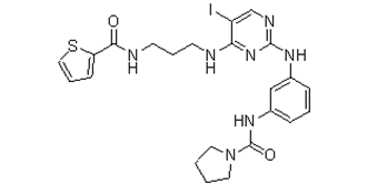
Formula | C23H26IN7O2S |
CAS No. | 702675-74-9 |
Storage | -20℃ for 3 years in powder form |
-80℃ for 2 years in solvent | |
Solubility (In vitro) | DMSO: 100 mg/mL (169.1 mM) |
Water: <1 mg/mL | |
Ethanol: <1 mg/mL | |
Solubility (In vivo) | 30% PEG400+0.5% Tween80+5% propylene glycol: 30mg/mL |
Synonyms | BX 795; BX-795; BX795; N-[3-[[5-Iodo-4-[[3-[(2-thienylcarbonyl)amino]propyl]amino]-2-pyrimidinyl]amino]phenyl]-1-pyrrolidinecarboxamide |
In Vitro | In vitro activity: BX-795 effectively blocks PDK1 activity in PC-3 cells, as shown by their ability to block phosphorylation of S6K1, Akt, PKCδ, and GSK3β. BX-795 potently inhibits tumor cell growth on plastic with IC50 of 1.6, 1.4, and 1.9 μM for MDA-468, HCT-116 and MiaPaca cells, respectively. In soft agar, BX-795 displays higher growth inhibition with IC50 of 0.72, and 0.25 μM for MDA-468, and PC-3 cells, respectively. In addition, BX-795, as an inhibitor of the TBK1/IKKe, blocks TBK1- and IKKε-mediated activation of IRF3 and production of IFN-β. In platelet physiological responses, BX795 produces inhibitory effect on 2-MeSADP-induced or collagen-induced aggregation, ATP secretion and thromboxane generation.
Kinase Assay: PDK1 was assayed in a direct kinase assay and a coupled assay format measuring PDK1- and PtdIns-3,4-P2-mediated activation of AKT2. For the coupled assay, the final assay mixture (60 μL) contained: 15 mM MOPS, pH 7.2, 1 mg/mL bovine serum albumin, 18 mM β-glycerol phosphate, 0.7 mM dithiothreitol, 3 mM EGTA, 10 mM MgOAc, 7.5 μM ATP, 0.2 μCi of [γ-33P]ATP, 7.5 μM biotinylated peptide substrate (biotin-ARRRDGGGAQPFRPRAATF), 0.5 μL of PtdIns-3,4-P2-containing phospholipid vesicles, 60 pg of purified recombinant human PDK1, and 172 ng of purified recombinant human AKT2. After incubation for 2 hrs at room temperature, the biotin-labeled peptide was captured from 10 μL of the assay mixture on streptavidin-coated SPA beads, and product formation was measured by scintillation proximity in a Wallac MicroBeta counter. The product formed was proportional to the time of incubation and to the amount of PDK1 and inactive AKT2 added. PDK1 was added at suboptimal levels so that the assay could sensitively detect inhibitors of AKT2 activation as well as direct inhibitors of PDK1 or AKT2. To measure PDK1 activity directly, the final assay mixture (60 μL) contained 50 mM Tris-HCl, pH 7.5, 0.1 mM EGTA, 0.1 mM EDTA, 0.1% β-mercaptoethanol, 1 mg/mL bovine serum albumin, 10 mM MgOAc, 10 μM ATP, 0.2 μCi of [γ-33P]ATP, 7.5 μM substrate peptide (H2N-ARRRGVTTKTFCGT), and 60 ng of purified recombinant human PDK1. After 4 hrs at room temperature, we added 25 mM EDTA and spotted a portion of the reaction mixture on Whatman P81 phosphocellulose paper. The filter paper was washed three times with 0.75% phosphoric acid and once with acetone. After drying, the filter-bound labeled peptide was quantified using a Fuji phosphorimager.
Cell Assay: Cells seeded at a low density (1,500–3,000 cells/well, 0.1 mL/well, 96-well plates) are incubated overnight. Compound treatments are made by adding 10 μL/well of the compound in 1% dimethyl sulfoxide and growth medium (final concentration of dimethyl sulfoxide, 0.1%), followed by brief shaking. Treated cells are incubated for 72 hours, and viability is measured by the addition of 10 μL of the metabolic dye WST-1. The WST-1 signal is read in a plate reader at 450 nm, and a no cell, or zero time cell, background is subtracted to calculate the net signal. Results are reported as the average ± S.E. of two or more replicates. |
In Vivo | NA |
Animal model | NA |
Formulation & Dosage | NA |
References | J Biol Chem. 2005 May 20;280(20):19867-74; J Biol Chem. 2009 May 22;284(21):14136-46; Thromb Haemost. 2014 Mar 3;111(3):508-17. |
 m.cnreagent.com