| CAS NO: | 154447-35-5 |
| 规格: | ≥98% |
| 包装 | 价格(元) |
| 5mg | 电议 |
| 50mg | 电议 |
| 100mg | 电议 |
| 250mg | 电议 |
| 500mg | 电议 |
| 1g | 电议 |
Molecular Weight (MW) | 281.31 |
Formula | C17H15NO3 |
CAS No. | 154447-35-5 (NU-7026); |
Storage | -20℃ for 3 years in powder form |
-80℃ for 2 years in solvent | |
Solubility (In vitro) | DMSO: 1 mg/mL (3.55 mM) |
Water: <1 mg/mL | |
Ethanol: <1 mg/mL | |
Solubility (In vivo) | 1% DMSO+30% polyethylene glycol+1% Tween 80: 30 mg/mL |
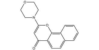
Synonyms | DNA-PK Inhibitor II; LY-293646; LY 293646; LY293646; NU7026; NU-7026; NU 7026. Chemical Name: 2-morpholino-4H-benzo[h]chromen-4-one InChi Key: KKTZALUTXUZPSN-UHFFFAOYSA-N InChi Code: InChI=1S/C17H15NO3/c19-15-11-16(18-7-9-20-10-8-18)21-17-13-4-2-1-3-12(13)5-6-14(15)17/h1-6,11H,7-10H2 SMILES Code: O=C1C=C(N2CCOCC2)OC3=C1C=CC4=CC=CC=C43 |
In Vitro | In vitro activity: NU7026 potentiates ionizing radiation induced cytotoxicity in a concentration-dependent manner in V3YAC and PARP-1+/+ cells. NU7026 completely abolishes potentially lethal damage recovery in growth-arrested cells. NU7026 inhibits DNA DSB repair by 56% in the V3YAC cell line. NU7026 (10 μM) potentiates the growth inhibitory effects of idarubicin, daunorubicin, doxorubicin, etoposide, mAMSA, and mitoxantrone with PF50 values ranging from approximately 19 for mAMSA to approximately 2 for idarubicin in K562 cells. NU7026 (10 μM) also potentiates the growth inhibitory effect of etoposide in this leukemia cell line with a PF50 value of 10.53. NU7026 (10 μM) enhances the etoposide-induced cell cycle G2 blockade in K562 cells. NU7026 potentiates topo II poisons involves inhibition of nonhomologous end joining and a G2/M checkpoint arrest. NU7026 (10 μM) exposure of 4 h in combination with 3 Gy radiation is required for a significant radiosensitisation effect in CH1 human ovarian cancer cells. NU7026 (< 10 μM) plus chlorambucil has synergistic cytotoxic activity at nontoxic doses of NU7026 in a CLL cell line (I83) and in primary CLL-lymphocytes. NU7026 (10 μM) increases chlorambucil-induced G(2)/M arrest in I83 cells. NU7026 (10 μM) enhances chlorambucil -induced γH2AX throughout the cell cycle in the I83 cell line. NU7026 (10 μM) Increases chlorambucil-Induced apoptosis in the I83 cell line. NU7026 (55 μM) results in a dramatic induction of telomere fusion in p53 null MEFs and significantly fewer telomere fusions in p53 and ligase IV double null MEFs
Kinase Assay: Mammalian DNA-PK (500 ng/μL) is isolated from HeLa cell nuclear extract after chromatography using Q-Sepharose, S-Sepharose, and Heparin agarose. DNA-PK (250 ng) activity is measured at 30°C, in a final volume of 40 μL, in buffer containing 25 mM HEPES (pH 7.4), 12.5 mM MgCl2, 50 mM KCl, 1 mM DTT, 10% v/v Glycerol, 0.1% w/v NP-40, and 1 mg of the substrate GST-p53N66 (the NH2-terminal 66 amino acid residues of human wild-type p53 fused to glutathione S-transferase) in polypropylene 96-well plates. To the assay mix, varying concentrations of inhibitor (in DMSO at a final concentration of 1% v/v) are added. After 10 min of incubation, ATP is added to give a final concentration of 50 μM, along with a 30-mer double-stranded DNA oligonucleotide (final concentration of 0.5 ng/mL), to initiate the reaction. After 1 h with shaking, 150 μL of PBS are added to the reaction, and 5 μL are then transferred to a 96-well opaque white plate containing 45 μL of PBS per well, where the GSTp53N66 substrate is allowed to bind to the wells for 1 h. To detect the phosphorylation event on the serine 15 residue of p53 elicited by DNA-PK, a p53 phosphoserine-15 antibody is used in a basic ELISA procedure. An antirabbit horseradish peroxidase-conjugated secondary antibody is then used in the ELISA before the addition of chemiluminescence reagent to detect the signal as measured by chemiluminescent counting via a TopCount NXT.
Cell Assay: I83 cells are plated in RPMI 1640 medium with 10% FBS (1.5×10⁵ cells/mL) and treated with vehicle (DMSO), 5 μM CLB, CLB IC50, 10 μM NU7026, or the combination of both drugs for 0, 6, 24, and 48 h. Cell cycle distribution, apoptosis, DNA-PK phosphorylation, and γH2AX determination are determined, and they are expressed as a percentage of cells in each phase of the cycle. DNA content is analyzed with a FACSCalibur flow cytometer equipped with CellQuest software. |
In Vivo | NU7026 (20mg/kg, i.v.) undergoes rapid plasma clearance (0.108/hour) in mice and this is largely attributed to extensive metabolism. Bioavailability following interperitoneal (i.p.) and p.o. administration of NU7026 at dose of 20 mg/kg is 20 and 15%, respectively. |
Animal model | Female BALB/c mice |
Formulation & Dosage | Dissolved in 10% DMSO and 5% Tween 20 in saline; 25 mg/kg; i.p. injection or oral administration |
References | Blood. 2004 Jun 15;103(12):4659-65; Br J Cancer. 2005 Oct 31;93(9):1011-8; Cancer Res. 2009 Mar 1;69(5):2100-7; Cancer Res. 2003 Sep 15;63(18):6008-15. |
 m.cnreagent.com