| CAS NO: | 1346574-57-9 |
| 规格: | ≥98% |
| 包装 | 价格(元) |
| 50mg | 电议 |
| 100mg | 电议 |
| 250mg | 电议 |
| 500mg | 电议 |
| Molecular Weight (MW) | 526.67 |
|---|---|
| Formula | C31H38N6O2 |
| CAS No. | 1346574-57-9 |
| Storage | -20℃ for 3 years in powder form |
| -80℃ for 2 years in solvent | |
| Solubility (In vitro) | DMSO: 3 mg/mL (5.7 mM) |
| Water: <1 mg/mL | |
| Ethanol: <1 mg/mL | |
| Solubility (In vivo) | 4% DMSO+corn oil: 0.5 mg/mL |
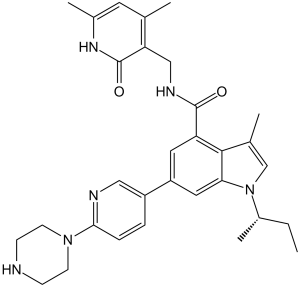
| Synonyms | GSK-2816126; GSK 2816126; GSK2816126; GSK126; GSK-126; GSK 126 Chemical Name: (S)-1-(sec-butyl)-N-((4,6-dimethyl-2-oxo-1,2-dihydropyridin-3-yl)methyl)-3-methyl-6-(6-(piperazin-1-yl)pyridin-3-yl)-1H-indole-4-carboxamide InChi Key: FKSFKBQGSFSOSM-QFIPXVFZSA-N InChi Code: InChI=1S/C31H38N6O2/c1-6-22(5)37-18-20(3)29-25(30(38)34-17-26-19(2)13-21(4)35-31(26)39)14-24(15-27(29)37)23-7-8-28(33-16-23)36-11-9-32-10-12-36/h7-8,13-16,18,22,32H,6,9-12,17H2,1-5H3,(H,34,38)(H,35,39)/t22-/m0/s1 SMILES Code: O=C(C1=CC(C2=CC=C(N3CCNCC3)N=C2)=CC4=C1C(C)=CN4[C@@H](C)CC)NCC5=C(C)C=C(C)NC5=O |
| In Vitro | In vitro activity: In vitro, GSK126 most potently inhibits H3K27me3, followed by H3K27me2 in both EZH2 wild-type and mutant DLBCL cell lines. GSK126 also effectively inhibits the proliferation of EZH2 mutant DLBCL cell lines, and induces transcriptional activation of EZH2 target genes in sensitive cell lines. In A687V EZH2-mutant cells, GSK126 treatment results in a global decrease in H3K27me3, robust gene activation, caspase activation, and decreased proliferation. In parental H2087 cells, GSK126 inhibits the expression of VEGF-A and phosphorylated Ser(473)-AKT, and thus causes the inhibition of cell proliferation, migration and metastasis. Kinase Assay: The five-member PRC2 complex (Flag–EZH2, EED, SUZ12, AEBP2, RbAp48) containing either wild-type or mutant EZH2 is prepared. GSK126 is dissolved in DMSO and tested at concentrations of 0.6 nM to 300 nM with a final DMSO concentration of 2.5%. In contrast to wild-type EZH2 which prefers H3K27me0 as a substrate in vitro, EZH2 Y641 mutants prefer H3K27me2 and have little activity with H3K27me0 or H3K27me1. The A677G mutant is distinct from both the wild-type and Y641 mutant forms of EZH2 in that it efficiently methylates H3K27me0, H3K27me1, and H3K27me2; therefore, histone H3 peptides (residues 21–44; 10 μM final) with either K27me0 (wild type, A677G EZH2), K27me1 (A677G EZH2), or K27me2 (A677G, Y641N, Y641C, Y641H, Y641S and Y641F EZH2) are used as methyltransferase substrates. GSK126 is added to plates followed by addition of 6 nM EZH2 complex and peptide. As the potency of GSK126 is at or near the tight binding limit of an assay run at [SAM] = Km, IC50 values are measured at a high concentration of the competitive substrate SAM relative to its Km (7.5 μM SAM where the SAM Km is 0.3 μM). Under these conditions, the contribution from the enzyme concentration becomes relatively small and accurate estimates of Ki can be calculated. Reactions are initiated with [3H]-SAM, incubated for 30 min, quenched with the addition of 500-fold excess unlabelled SAM, and the methylated product peptide is captured on phosphocellulose filters according to the vendor supplied protocol for MSPH Multiscreen plates. Plates are read on a TopCount after adding 20 μL of Microscint-20 cocktail. Apparent Ki values are calculated using the Cheng–Prusoff relationship for a competitive inhibitor. IC50=Ki (1+[S]/Km)+[E]/2, where E is the enzyme and S is the substrate. Cell Assay: The optimal cell seeding is determined empirically for all cell lines by examining the growth of a wide range of seeding densities in a 384-well format to identify conditions that permitted proliferation for 6 days. Cells (46 lymphoma cell lines) are then plated at the optimal seeding density 24 h before treatment (in duplicate) with a 20-point two fold dilution series of GSK126 or 0.15% DMSO. Plates are incubated for 6 days at 37°C in 5% CO2. Cells are then lysed with CellTiter-Glo (CTG) and chemiluminescent signal is detected with a TECAN Safire2 microplate reader. In addition, an untreated plate of cells is harvested at the time of compound addition (T0) to quantify the starting number of cells. CTG values obtained after the 6 day treatment are expressed as a percent of the T0 value and plotted against compound concentration. Data are fit with a four-parameter equation to generate a concentration response curve and the concentration of GSK126 required to inhibit 50% of growth (growth IC50) is determined. |
|---|---|
| In Vivo | In mice bearing KARPAS-422 and Pfeiffer xenografts, GSK126 (150 mg/kg/d, i.p.) decreases global H3K27me3, increases gene expression, and thus causes marked tumour regression. |
| Animal model | Female beige SCID mice bearing Pfeiffer or KARPAS-422 tumors |
| Formulation & Dosage | Dissolved in 20% captisol; 150 mg/kg; i.p. |
| References | Nature. 2012 Dec 6;492(7427):108-12; Mol Cancer Ther. 2014 Dec;13(12):3062-73. |
 m.cnreagent.com