| CAS NO: | 128794-94-5 |
| 规格: | ≥98% |
| 包装 | 价格(元) |
| 250mg | 电议 |
| 500mg | 电议 |
| 1g | 电议 |
| 2g | 电议 |
| 5g | 电议 |
| 10g | 电议 |
| Molecular Weight (MW) | 433.49 |
|---|---|
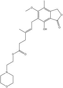
| Formula | C23H31NO7 |
| CAS No. | 128794-94-5 |
| Storage | -20℃ for 3 years in powder form |
| -80℃ for 2 years in solvent | |
| Solubility (In vitro) | DMSO: 86 mg/mL (198.4 mM) |
| Water: <1 mg/mL | |
| Ethanol: <1 mg/mL | |
| Solubility (In vivo) | 0.5% methylcellulose: 20 mg/mL |
| Synonyms | RS61443; Mycophenolic acid, Mycophenolate mofetil, Cellcept, Myfortic, RS-61443; Mycophenolate mofetil (free acid); |
| In Vitro | In vitro activity: Mycophenolate mofetil is an ester prodrug of the active immunosuppressant mycophenolic acid (MPA). The latter shows a noncompetitive, selective and reversible inhibition activity against inosine monophosphate dehydrogenase type I/II with IC50 of 39 nM and 27 nM, respectively. Moreover, MPA also produces the concentration-dependent inhibition of proliferation of ConA-stimulated T cells, LPS-stimulated B cells and alloantigen-specific T cells with IC50 of 100 nM, 120 nM, and 51 nM, respectively. Mycophenolate mofetil with high concentration of 10 μg/mL induces a strong apoptosis in microglial cell cultures and increases the number of activated caspase-3 immunoreactive apoptotic cells. In addition, Mycophenolate mofetil (1 μg/mL) strongly inhibits proliferation of both microglial cells and astrocytes. A recent study shows that Mycophenolate mofetil significantly attenuates the extent of neuronal cell death of organotypic hippocampal slice cultures after neuronal injury in a time-dependent manner. Kinase Assay: IMP dehydrogenase Types I and II are purified from E. coli expressing human enzymes. The assay is performed using a flat bottom, UV-transparent 96-well plate. The final 200 μL reaction mixture contained 0.1 M Tris, 0.1 M KCl, 3 mM EDTA pH 8.0, 2 mM DTT, and 40 nM of either IMP dehydrogenase Type I or Type II. The reaction is initiated by adding 400 μM NAD and 400 μM IMP, followed by incubation at 37 °C for 2.5 hours. The reaction rate of the conversion of NAD to NADH is then measured based on the increase in absorbance at 340 nm. The assays are also performed in the presence of 50% human serum to estimate serum protein binding by different IMP dehydrogenase inhibitors. Cell Assay: The spleens of male Lewis rats aged 8 weeks are aseptically removed and teased into single-cell suspensions, and the resulting splenocytes are suspended in RPMI1640 medium containing 10% fetal calf serum, 100 U/mL penicillin, and 100 μg/mL streptomycin. Assays are performed in flat-bottomed microtiter plates, with each well containing 150000 splenocytes in a total volume of 100 μL. Splenocytes are incubated in medium containing either 1 μg/mL Concanavalin A (ConA) or lipopolysaccharide (LPS) as T or B cell mitogen, respectively, along with various concentrations of MPA at 37 °C for 48 hours in a humidified atmosphere of 5% CO2-95% air. During the final 6 hours of incubation, cells are pulsed with 1 μCi of 3H-thymidine/well, and harvested onto pressed fiberglass in a cell harvester. The uptake of 3H-thymidine by proliferating cells is measured using a liquid scintillation counter. The in vitro immune response of alloantigen-specific T cells is evaluated as a proliferation response using one-way mixed lymphocyte reaction assay. Mesenteric lymph nodes cells from male Lewis rats aged 8 weeks and splenocytes from male ACI rats aged 8 weeks are used as responder and stimulator cells, respectively. Single-cell suspensions are prepared, and the responder cells are cultured with irradiated (20 Gy) stimulator cells in RPMI1640 supplemented with 10% fetal calf serum, 100 U/mL penicillin and 100 μg/mL streptomycin, in 96-well plates (U-bottom) and in the presence of various concentrations of MPA (total volume: 200 μL, 37 °C, 5% CO2 humidified atmosphere, 72 hours). As a negative control, responder cells are cultured with irradiated splenocytes of Lewis rats. The cell proliferation is quantified by the uptake of 3H-thymidine. |
|---|---|
| In Vivo | In an ACI-to-Lewis rat heterotopic cardiac transplant model, treatment of Mycophenolate mofetil at doses of 20 mg/kg and 40 mg/kg leads to a prolongation of graft survival, with median survival time (MST) of 14.5 days and 18.5 days, respectively. In bleomycin (BLM)-induced scleroderma mouse model, Mycophenolate mofetil reduces inflammatory-cell infiltration, tissue hydroxyproline content and dermal thickness. |
| Animal model | Lewis rats with abdominal vascularized heterotopic cardiac transplantation |
| Formulation & Dosage | Dissolved in 0.5% methylcellulose water; 40 mg/kg; i.v. administration |
| References | Int Immunopharmacol. 2010 Jan;10(1):91-7; Clin Exp Dermatol. 2012 Jan;37(1):48-54. |
 m.cnreagent.com