| CAS NO: | 587871-26-9 |
| 规格: | ≥98% |
| 包装 | 价格(元) |
| 5mg | 电议 |
| 10mg | 电议 |
| 25mg | 电议 |
| 50mg | 电议 |
| 100mg | 电议 |
| 250mg | 电议 |
| 500mg | 电议 |
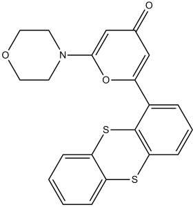
| Molecular Weight (MW) | 395.49 |
|---|---|
| Formula | C21H17NO3S2 |
| CAS No. | 587871-26-9 |
| Storage | -20℃ for 3 years in powder form |
| -80℃ for 2 years in solvent | |
| Solubility (In vitro) | DMSO: 33 mg/mL (83.4 mM) |
| Water: <1 mg/mL | |
| Ethanol: <1 mg/mL | |
| Solubility (In vivo) | 5% DMSO and 47.5% PEG300: 10 mg/mL |
| Synonyms | KU 55933; KU55933; KU-55933 |
| In Vitro | In vitro activity: KU-55933 inhibits DNA-PK and PI3K with IC50 of 2.5 μM and 16.6 μM, respectively. Besides, KU-55933 also prevents the activity of mTOR with IC50 of 9.3 μM. KU-55933 is active at the cellular level in ablating a well-characterized ATM-dependent phosphorylation event. KU-55933 has a dose-dependent effect in inhibiting this ATM-dependent phosphorylation event with IC50 of 300 nM. KU-58050 does not prevent the ATM-dependent phosphorylation of p53 serine 15 until a dose of 30 μM. Addition of KU-55933 has no appreciable effects on UV-induced phosphorylation of H2AX on serine 139, NBS1 on serine 343, CHK1 on serine 345, and SMC1 on serine 966. In stark contrast to the UV responses, KU-55933 ablates the ionizing radiation-induced phosphorylation of these ATM substrates. KU-55933 sensitizes HeLa cells to a range of ionizing radiation doses. KU-55933 inhibits the phosphorylation of Akt induced by growth factors in cancer cells. KU-55933 suppresses the proliferation of cancer cells. Furthermore, suppression of ATM by KU-55933 improves survival, probably via prevention of downstream activation of TAp63α. Kinase Assay: ATM for use in the in vitro assay is obtained from HeLa nuclear extract by immunoprecipitation with rabbit polyclonal antiserum raised to the COOH-terminal 400 amino acids of ATM in buffer containing 25 mM HEPES (pH 7.4), 2 mM MgCl2, 250 mM KCl, 500 μM EDTA, 100 μM Na3VO4, 10% v/v glycerol, and 0.1% v/v Igepal. ATM-antibody complexes are isolated from nuclear extract by incubating with protein A-Sepharose beads for 1 hour and then through centrifugation to recover the beads. In the well of a 96-well plate, ATM-containing Sepharose beads are incubated with 1 μg of substrate glutathione S-transferase–p53N66 (NH2-terminal 66 amino acids of p53 fused to glutathione S-transferase) in ATM assay buffer [25 mM HEPES (pH 7.4), 75 mM NaCl, 3 mM MgCl2, 2 mM MnCl2, 50 μM Na3VO4, 500 μM DTT, and 5% v/v glycerol] at 37 °C in the presence or absence of inhibitor. After 10 minutes with gentle shaking, ATP is added to a final concentration of 50 μM and the reaction continued at 37 °C for an additional 1 hour. The plate is centrifuged at 250 × g for 10 minutes (4 °C) to remove the ATM-containing beads, and the supernatant is removed and transferred to a white opaque 96-well plate and incubated at room temperature for 1.5 hours to allow glutathione S-transferase-p53N66 binding. This plate is then washed with PBS, blotted dry, and analyzed by a standard ELISA technique with a phospho-serine 15 p53 antibody. The detection of phosphorylated glutathione S-transferase-p53N66 substrate is performed in combination with a goat antimouse horseradish peroxidase-conjugated secondary antibody. Enhanced chemiluminescence solution is used to produce a signal and chemiluminescent detection is carried out Cell Assay: U2OS cells are exposed to ionizing radiation (3, 5, or 15 Gy) or UV (5 or 50 J/m2) and the ATM response determined by Western blot analysis of p53 serine 15 phosphorylation and stabilization of wild-type p53. Whole cell extracts are obtained from each time point, proteins separated by SDS-PAGE, and the ATM-specific increase in phosphorylated serine 15 measured with a p53 phospho-serine 15 specific antibody. Overall p53 stabilization with time is also observed with a p53-specific antibody (DO-1). Similarly, for studying ATM-dependent phosphorylations on H2AX, CHK1, NBS1, and SMC1, the following antibodies are used: CHK1 phospho-serine 345 and NBS1 phospho-serine 343 antibodies. Histone H2A (H-124) and CHK1 antibodies are also used, as well as SMC1 and SMC1 phospho-serine 966 antibodies. For determination of a cellular IC50 for KU-55933, the peak response time for p53 serine 15 phosphorylation of 2 hours is used to monitor inhibition of ATM. KU-55933 is titrated onto cells and preincubated for 1 hour before ionizing radiation. Using scanning densitometry, the percentage inhibition relative to vehicle control is calculated, and the IC50 value is calculated as for the in vitro determinations. |
|---|---|
| In Vivo | Suppression of ATM-dependent STAT3 activation by KU-55933 enhances TRAIL-mediated apoptosis through up-regulation of surface DR5 expression, whereas suppression of both STAT3 and NF-κB appeares to be involved in down-regulation of cFLIP accompanied by an additional increase in apoptotic levels. The ATM inhibitor KU-55933 affectes TRAIL-mediated apoptosis more strongly than the JAK2 inhibitor, AG490, or overexpression of STAT3β. |
| Animal model | |
| Formulation & Dosage | |
| References | Cancer Res. 2009 Apr 15;69(8):3510-9; Mol Cancer Ther. 2010 Jan;9(1):113-25. |
 m.cnreagent.com