| CAS NO: | 186497-07-4 |
| 规格: | ≥98% |
| 包装 | 价格(元) |
| 5mg | 电议 |
| 10mg | 电议 |
| 25mg | 电议 |
| 50mg | 电议 |
| 100mg | 电议 |
| 250mg | 电议 |
| 500mg | 电议 |
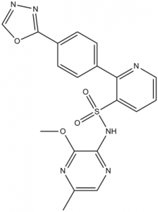
| Molecular Weight (MW) | 424.43 |
|---|---|
| Formula | C19H16N6O4S |
| CAS No. | 186497-07-4 |
| Storage | -20℃ for 3 years in powder form |
| -80℃ for 2 years in solvent | |
| Solubility (In vitro) | DMSO: 24 mg/mL (56.5 mM) |
| Water: <1 mg/mL | |
| Ethanol:<1 mg/mL | |
| Solubility (In vivo) | 1% DMSO+30% polyethylene glycol+1% Tween 80: 30 mg/mL |
| Synonyms | ZD4054; ZD-4054; ZD 4054 |
| In Vitro | In vitro activity: As Zibotentan specifically inhibits ETA-mediated antiapoptotic effects, but not ETB-mediated proapoptotic effects in human and rat smooth muscle cells, Zibotentan binds to endothelin A receptor (ETA) with high affinity with Ki of 13 nM, and has no affinity for endothelin B receptor (ETB) with IC50 of>10 μM. Zibotentan treatment at 1 μM inhibits ET-1 induced mitogenic activity in ovarian carcinoma cell lines HEY and OVCA 433 secreting ET-1 and expressing ETA and ETB mRNA. ZD4054 (1 μM) inhibits ET-1 induced EGFR transactivation in HEY and OVCA 433 cells. Zibotentan (1 μM) reverts ET-1 mediated epithelial-mesenchymal transition (EMT), by enhancing E-cadherin expression and promoter activity, and inhibiting vascular endothelial growth factor (VEGF) secretion and invasiveness in HEY and OVCA 433 cells. Zibotentan also potently inhibits the basal and ET-1 induced cell proliferation in SKOV-3 and A-2780 cells, associated with the inhibition of AKT and p42/44MAPK phosphorylation, and with increased apoptosis through the inhibition of bcl-2 and activation of caspase-3 and poly(ADP-ribose) polymerase proteins. Kinase Assay: The inhibition by Zibotentan (varying concentrations) of 125iodine-ET-1 binding to cloned human ETA is assessed using standard radioligand-binding techniques. Human recombinant ETA is expressed in mouse erythroleukaemic cells, and cell membranes prepared for competitive binding studies using 125iodine-ET-1 as the radioligand. Incubations are carried out in triplicate in the presence of Zibotentan, 100 pM to 100 μM in half-log increments, and inhibition of ET-1 binding is expressed as the geometric mean pIC50 value (concentration to inhibit 50% of binding) with a 95% confidence interval (CI). The affinity of Zibotentan for cloned human ETA is also assessed using the equation of Cheng and Prusoff to determine the equilibrium dissociation constant (Ki) in a further receptor-binding screen utilizing a greater number of concentration-response curves determined in three separate studies. Cell Assay: Cells (HEY and OVCA 433) are serum starved by incubation for 24 hours in serum-free DMEM before exposed to Zibotentan for 48 hours. After the treatment, cells are lysed and the supernatant is recovered and assayed for histone-associated DNA fragments, at 405 nm by the use of a microplate reader. For detection of early apoptotic events, floating and adherent cells are collected. Cells are double stained with FITC-conjugated Annexin V and propidium iodide using the Vybrant Apoptosis Kit and are immediately analyzed by cytofluorometric analysis. |
|---|---|
| In Vivo | Administration of Zibotentan at 10 mg/kg/day for 21 days potently inhibits the growth of HEY ovarian carcinoma xenografts in mice by 69% with no associated toxicity, which is in association with the blocking of cell proliferation evaluated by 37% inhibition of the Ki-67 expression, and the 62% inhibition of tumor-induced vascularization. Consistently, Zibotentan treatment significantly inhibits the expression of matrix metalloproteinase-2 (MMP-2) and VEGF, as well as the activation of p42/44 MAPK and EGFR, and potently enhances the expression of E-cadherin. |
| Animal model | Female athymic (nu+/nu+) mice bearing established HEY human ovarian carcinoma xenografts |
| Formulation & Dosage | Dissolved in DMSO, and diluted in PBS; 10 mg/kg; i.p. injection |
| References | Br J Cancer. 2005 Jun 20;92(12):2148-52; Exp Biol Med (Maywood). 2006 Jun;231(6):1132-5; Cancer Res. 2007 Jul 1;67(13):6351-9. |
 m.cnreagent.com