| CAS NO: | 790299-79-5 |
| 规格: | ≥98% |
| 包装 | 价格(元) |
| 50mg | 电议 |
| 100mg | 电议 |
| 250mg | 电议 |
| 500mg | 电议 |
| 1g | 电议 |
| 2g | 电议 |
| Molecular Weight (MW) | 498.64 |
|---|---|
| Formula | C28H30N6OS |
| CAS No. | 790299-79-5 |
| Storage | -20℃ for 3 years in powder form |
| -80℃ for 2 years in solvent | |
| Solubility (In vitro) | DMSO: 100 mg/mL (200.5 mM) |
| Water: <1 mg/mL | |
| Ethanol: 4 mg/mL (8.0 mM) | |
| Solubility (In vivo) | 4% DMSO+30% PEG 300+5% Tween 80+ddH2O: 30mg/mL |
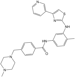
| Synonyms | AB-1010; AB 1010; AB1010; Masitinib. Brand name: Kinavet; Masivet. Chemical Name: 4-[(4-methylpiperazin-1-yl)methyl]-N-(4-methyl-3-{[4-(pyridin-3-yl)-1,3-thiazol-2-yl]amino}phenyl)benzamide InChi Key: WJEOLQLKVOPQFV-UHFFFAOYSA-N InChi Code: InChI=1S/C28H30N6OS/c1-20-5-10-24(16-25(20)31-28-32-26(19-36-28)23-4-3-11-29-17-23)30-27(35)22-8-6-21(7-9-22)18-34-14-12-33(2)13-15-34/h3-11,16-17,19H,12-15,18H2,1-2H3,(H,30,35)(H,31,32) SMILES Code: O=C(NC1=CC=C(C)C(NC2=NC(C3=CC=CN=C3)=CS2)=C1)C4=CC=C(CN5CCN(C)CC5)C=C4 |
| In Vitro | In vitro activity: Masitinib is a competitive inhibitor against ATP at concentrations ≤500 nM. Masitinib also potently inhibits recombinant PDGFR and the intracellular kinase Lyn, and to a lesser extent, fibroblast growth factor receptor 3. In contrast, Masitinib demonstrates weak inhibition of Abl and c-Fms. Masitinib more strongly inhibits degranulation, cytokine production, and bone marrow mast cell migration than imatinib. In Ba/F3 cells expressing human wild-type Kit, Masitinib inhibits SCF (stem cell factor)-induced cell proliferation with an IC50 of 150 nM, while the IC50 for inhibition of IL-3-stimulated proliferation is at approximately>10 μM. In Ba/F3 cells expressing PDGFRα, Masitinib inhibits PDGF-BB-stimulated proliferation and PDGFRα tyrosine phosphorylation with IC50 of 300 nM. Masitinib also causes inhibition of SCF-stimulated tyrosine phosphorylation of human Kit in mastocytoma cell-lines and BMMC. Masitinib inhibits Kit gain-of-function mutants, including V559D mutant and Δ27 mouse mutant with IC50 of 3 and 5 nM in Ba/F3 cells. Masitinib inhibits the cell proliferation of mastocytoma cell lines including HMC-1α155 and FMA3 with IC50 of 10 and 30 nM, respectively. Masitinib inhibits cell growth and PDGFR phosphorylation in two novel ISS cell lines, which suggest that Masitinib displays activity against both primary and metastatic ISS cell line and may aid in the clinical management of ISS. Kinase Assay: A 96-well microtitre plateis coated overnight with 0.25 mg/ml poly(Glu,Tyr 4:1), rinsed twice with 250 μL of washing buffer (10 mM phosphate-buffered saline [pH 7.4] and 0.05% Tween 20) and dried for 2 hours at room temperature. Assays are performed at room temperature with a final volume of 50 μL in kinase buffer (10 mM MgCl2, 1 mM MnCl2, 1 mM sodium orthovanadate, 20 mM HEPES, pH 7.8) containing ATP at a concentration of at least twice the Km for each enzyme and an appropriate amount of recombinant enzyme to ensure a linear reaction rate. Reactions are initiated upon introduction of the enzyme and terminated with the addition of one reaction volume (50 μL) of 100 mM EDTA per 5 M urea mix. Plates are washed three times and incubated with 1:30,000 horseradish peroxidase-conjugated anti-phosphotyrosine monoclonal antibody, then washed three times and incubated with tetramethylbenzidine. The final reaction product is quantified by spectrophotometry at 450 nm. Cell Assay: For the assay of Ba/F3 cell proliferation, microtitre plates are seeded with a total of 104 cells/well in 100 μL of RPMI 1640 medium with 10% foetal bovine serum at 37 °C. These are supplemented, or not, with either 0.1% conditioned medium from X63-IL-3 cells or 250 ng/mL murine SCF. The murine SCF, which activates Kit, is purified from the conditioned medium of SCF-producing CHO cells. Cells are grown for 48 hours at 37 °C with Masitinib and then incubated with 10 μL/well of WST-1 reagent for 3 hours at 37 °C. The amount of formazan dye formed is quantified by its absorbance at 450 nm using a scanning multiwell spectrophotometer. A blank well without cells is used as a background control for the spectrophotometer. Cells: Ba/F3 cells expressing wild-type or mutant human Kit, HMC1, HMC-1α155 |
|---|---|
| In Vivo | Masitinib inhibits tumour growth and increases the median survival time in Δ27-expressing Ba/F3 tumor models at 30 mg/kg, without cardiotoxicity or genotoxicity. Masitinib (12.5 mg/kg/d PO) increases overall TTP (time-to-tumor progression) compared with placebo in dogs. The combination of masitinib/gemcitabine shows synergy in vitro on proliferation of gemcitabine-refractory cell lines Mia Paca2 and Panc1, and to a lesser extent on Mia Paca-2 pancreatic tumours in NogCID mice |
| Animal model | Female MBRI Nu/Nu mice bearing a/F3 Δ27 tumour model |
| Formulation & Dosage | Dissolved in DMSO; 30 mg/kg (i.p.) or 10, 30, or 45 mg/kg (p.o.).; i.p. or oral gavage |
| References | PLoS One. 2009; 4(9): e7258; PLoS One. 2010 Mar 4;5(3):e9430 |
 m.cnreagent.com