| CAS NO: | 877399-52-5 |
| 规格: | ≥98% |
| 包装 | 价格(元) |
| 25mg | 电议 |
| 50mg | 电议 |
| 100mg | 电议 |
| 250mg | 电议 |
| 500mg | 电议 |
| 1g | 电议 |
| 5g | 电议 |
| Molecular Weight (MW) | 450.34 |
|---|---|
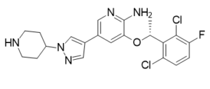
| Formula | C21H22Cl2FN5O |
| CAS No. | 877399-52-5 (free base); |
| Storage | -20℃ for 3 years in powder form |
| -80℃ for 2 years in solvent | |
| Solubility (In vitro) | DMSO: 9 mg/mL (20 mM) |
| Water: <1 mg/mL | |
| Ethanol: <1 mg/mL | |
| Solubility (In vivo) | 5% DMSO+30% PEG 300+dd H2O: 5 mg/mL |
| Synonyms | PF-2341066; PF02341066; PF 2341066; Crizotinib; PF-02341066; PF 02341066; PF2341066; US trade name: Xalkori Chemical Name: (R)-3-(1-(2,6-dichloro-3-fluorophenyl)ethoxy)-5-(1-(piperidin-4-yl)-1H-pyrazol-4-yl)pyridin-2-amine InChi Key: KTEIFNKAUNYNJU-GFCCVEGCSA-N InChi Code: InChI=1S/C21H22Cl2FN5O/c1-12(19-16(22)2-3-17(24)20(19)23)30-18-8-13(9-27-21(18)25)14-10-28-29(11-14)15-4-6-26-7-5-15/h2-3,8-12,15,26H,4-7H2,1H3,(H2,25,27)/t12-/m1/s1 SMILES Code: NC1=NC=C(C2=CN(C3CCNCC3)N=C2)C=C1O[C@@H](C4=C(Cl)C=CC(F)=C4Cl)C |
| In Vitro | In vitro activity: PF-2341066 displays similar potency against c-Met phosphorylation in mIMCD3 mouse or MDCK canine epithelial cells with IC50 of 5 nM and 20 nM, respectivly. PF-2341066 shows improved or similar activity against NIH3T3 cells engineered to express c-Met ATP-binding site mutants V1092I or H1094R or the P-loop mutant M1250T with IC50 of 19 nM, 2 nM and 15 nM, respectively, compared with NIH3T3 cells expressing wild-type receptor with IC50 of 13 nM. In contrast, a marked shift in potency of PF-2341066 is observed against cells engineered to express c-Met activation loop mutants Y1230C and Y1235D with IC50 of 127 nM and 92 nM, respectively, compared with wild-type receptor. PF-2341066 also potently prevents the phosphorylation of c-Met in NCI-H69 and HOP92 cells, with IC50 of 13 nM and 16 nM, respectively, which express the endogenous c-Met variants R988C and T1010I, respectively. PF-2341066 is>1,000-fold selective for the VEGFR2 and PDGFRβ RTKs,>250-fold selective for IRK and Lck, and ~40- to 60-fold selective for Tie2, TrkA, and TrkB, all compared with c-Met. PF-2341066 is 20- to 30-fold selective for RON and Axl RTKs. In contrast, PF-2341066 shows a near-equivalent IC50 of 24 nM against the nucleophosmin (NPM)-anaplastic lymphoma kinase (ALK) oncogenic fusion variant of the ALK RTK expressed by the KARPAS299 human anaplastic large cell lymphoma (ALCL) cell line. PF-2341066 inhibits c-Met–dependent neoplastic phenotypes of cancer cells and angiogenic phenotypes of endothelial cells. PF-2341066 suppresses human GTL-16 gastric carcinoma cell growth with IC50 of 9.7 nM. PF-2341066 induces apoptosis in GTL-16 cells with IC50 of 8.4 nM. PF-2341066 inhibits HGF-stimulated human NCI-H441 lung carcinoma cell migration and invasion with IC50 of 11 nM and 6.1 nM, respectively. PF-2341066 inhibits MDCK cell scattering with IC50 of 16 nM. PF-2341066 prevents HGF-stimulated c-Met phosphorylation, cell survival, and Matrigel invasion with IC50 of 11 nM, 14 nM and 35 nM, respectively. In addition, PF-2341066 prevents serum-stimulated HMVEC branching tubulogenesis (formation of vascular tubes) in fibrin gels. PF-2341066 also potently inhibits NPM-ALK phosphorylation in Karpas299 or SU-DHL-1 ALCL cells with an IC50 of 24 nM. PF-2341066 potently prevents cell proliferation, which is associated with G(1)-S-phase cell cycle arrest and induction of apoptosis in ALK-positive ALCL cells with IC50 of 30 nM, but not ALK-negative lymphoma cells. Besides, PF-2341066 prevents osteosarcoma behavior associated with primary tumor growth (i.e., proliferation and survival) as well as metastasis (eg, invasion and clonogenicity). Kinase Assay: Cells are seeded in 96-well plates in media supplemented with 10% fetal bovine serum (FBS) and transferred to serum-free media [with 0.04% bovine serum albumin (BSA)] after 24 h. In experiments investigating ligand-dependent RTK phosphorylation, corresponding growth factors are added for up to 20 min. After incubation of cells with PF-2341066 for 1 h and/or appropriate ligands for the designated times, cells are washed once with HBSS supplemented with 1 mM Na3VO4, and protein lysates are generated from cells. Subsequently, phosphorylation of selected protein kinases is assessed by a sandwich ELISA method using specific capture antibodies used to coat 96-well plates and a detection antibody specific for phosphorylated tyrosine residues. Antibody-coated plates are (a) incubated in the presence of protein lysates at 4°C overnight; (b) washed seven times in 1% Tween 20 in PBS; (c) incubated in a horseradish peroxidase–conjugated anti–total-phosphotyrosine (PY-20) antibody (1:500) for 30 min; (d) washed seven times again; (e) incubated in 3,3′,5,5′-tetramethyl benzidine peroxidase substrate to initiate a colorimetric reaction that is stopped by adding 0.09 N H2SO4; and (f) measured for absorbance in 450 nm using a spectrophotometer. Cell Assay: Cells (GTL-16 gastric carcinoma cells and T47D breast carcinoma cells) including GTL-16 gastric carcinoma cells and T47D breast carcinoma cells are seeded in 96-well plates in media supplemented with 10% fetal bovine serum (FBS) and transferred to serum-free media [with 0.04% bovine serum albumin (BSA)] after 24 hours. In experiments investigating ligand-dependent RTK phosphorylation, corresponding growth factors are added for up to 20 minutes. After incubation of cells with PF-2341066 for 1 hour and/or appropriate ligands for the designated times, cells are washed once with HBSS supplemented with 1 mM Na3VO4, and protein lysates are generated from cells. Subsequently, phosphorylation of selected protein kinases is assessed by a sandwich ELISA method using specific capture antibodies used to coat 96-well plates and a detection antibody specific for phosphorylated tyrosine residues. Antibody-coated plates are (a) incubated in the presence of protein lysates at 4 °C overnight; (b) washed seven times in 1% Tween 20 in PBS; (c) incubated in a horseradish peroxidase–conjugated anti–total-phosphotyrosine (PY-20) antibody (1:500) for 30 min; (d) washed seven times again; (e) incubated in 3,3′,5,5′-tetramethyl benzidine peroxidase substrate to initiate a colorimetric reaction that is stopped by adding 0.09 N H2SO4; and (f) measured for absorbance in 450 nm using a spectrophotometer. |
|---|---|
| In Vivo | In the GTL-16 model, PF-2341066 reveals the ability to cause marked regression of large established tumors (>600 mm3) in both the 50 mg/kg/day and 75 mg/kg/day treatment cohorts, with a 60% decrease in mean tumor volume over the 43-day administration schedule. In an another study, PF-2341066 displays the ability to completely inhibits GTL-16 tumor growth for>3 months, with only 1 of 12 mice exhibiting a significant increase in tumor growth over the 3-month treatment schedule at 50 mg/kg/day. In the NCI-H441 NSCLC model, a 43% decrease in mean tumor volume is observed at 50 mg/kg/day during the 38-day PF-2341066 administration cycle. In the Caki-1 RCC model, a 53% decrease in mean tumor volume is observed to be associated with decreased volume of each tumor by at least 30% at 50 mg/kg/day during the 33-day PF-2341066 administration cycle. PF-2341066 also reveals near-complete prevention of the growth of established tumors at 50 mg/kg/day in the U87MG glioblastoma or PC-3 prostate carcinoma xenograft models, with 97% or 84% inhibition on the final study day, respectively. In contrast, PF-2341066 p.o. given at 50 mg/kg/day does not significantly inhibit tumor growth in the MDA-MB-231 breast carcinoma model, or the DLD-1 colon carcinoma model. A significant dose-dependent reduction of CD31–positive endothelial cells is observed at 12.5 mg/kg/day, 25 mg/kg/day, and 50 mg/kg/day in GTL-16 tumors, indicating that inhibition of MVD shows a dose-dependent correlation to antitumor efficacy. PF-2341066 displays a significant dose-dependent reduction of human VEGFA and IL-8 plasma levels in both the GTL-16 and U87MG models. Marked inhibition of phosphorylated c-Met, Akt, Erk, PLCλ1, and STAT5 levels is observed in GTL-16 tumors following p.o. administration of PF-2341066. P.o. administration of PF-2341066 to severe combined immunodeficient-Beige mice bearing Karpas299 ALCL tumor xenografts leads to dose-dependent antitumor efficacy with complete regression of all tumors at the 100 mg/kg/d dose within 15 days of initial compound administration. In addition, inhibition of key NPM-ALK signaling mediators, including phospholipase C-gamma, signal transducers and activators of transcription 3, extracellular signal-regulated kinases, and Akt by PF-2341066 are observed at concentrations or dose levels, which correlated with inhibition of NPM-ALK phosphorylation and function. PF-2341066 prevents osteosarcoma behavior associated with primary tumor growth (eg, proliferation and survival) as well as metastasis (eg, invasion and clonogenicity). In nude mice treated with PF-2341066 via oral gavage, the growth and associated osteolysis and extracortical bone matrix formation of osteosarcoma xenografts are prevented by PF-2341066. Treatment of c-MET-amplified GTL-16 xenografts with 50 mg/kg PF-2341066 elicits tumor regression that is associated with a slow reduction in 18F-FDG uptake and decreases expression of the glucose transporter 1, GLUT-1. |
| Animal model | Female SCID mice (5–6 weeks) |
| Formulation & Dosage | 25 (s.c.), 50 mg/kg (p.o.); s.c. or oral gavage |
| References | Cancer Res. 2007 May 1;67(9):4408-17; Mol Cancer Ther. 2007 Dec;6(12 Pt 1):3314-22; J Nucl Med. 2011 Aug;52(8):1261-7. |
 m.cnreagent.com