| CAS NO: | 91396-88-2 |
| 规格: | ≥98% |
| 包装 | 价格(元) |
| 5mg | 电议 |
| 10mg | 电议 |
| 25mg | 电议 |
| 50mg | 电议 |
| 100mg | 电议 |
| 250mg | 电议 |
| 500mg | 电议 |
| 1g | 电议 |
| Molecular Weight (MW) | 213.24 |
|---|---|
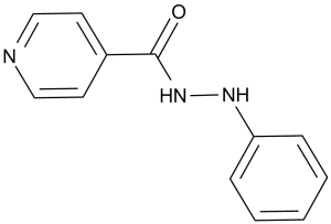
| Formula | C12H11N3O |
| CAS No. | 91396-88-2 |
| Storage | -20℃ for 3 years in powder form |
| -80℃ for 2 years in solvent | |
| Solubility (In vitro) | DMSO: 43 mg/mL (201.7 mM) |
| Water: <1 mg/mL | |
| Ethanol: <1 mg/mL | |
| Other info | Chemical Name: N'-phenylisonicotinohydrazide InChi Key: HUDWXDLBWRHCKO-UHFFFAOYSA-N InChi Code: InChI=1S/C12H11N3O/c16-12(10-6-8-13-9-7-10)15-14-11-4-2-1-3-5-11/h1-9,14H,(H,15,16) SMILES Code: O=C(NNC1=CC=CC=C1)C2=CC=NC=C2 |
| Synonyms | NSC 14613; PluriSln #1; NSC-14613; NSC14613; PluriSln-1 |
| In Vitro | In vitro activity: PluriSIns-1 has a robust, rapid, and selective cytotoxic effect toward hPSCs. Apoptosis is the central cell death mechanism activated by PluriSIn #1. PluriSIn #1 leads to ER stress in hPSCs. PluriSIn #1 (20 μM) induces ~30% decrease in protein synthesis in hPSCs. PluriSIns-1 exposure induces a ~65% decrease in stearoyl-coA desaturase (SCD1) activity in hPSCs. PluriSIns-1 (20 μM) prevents teratoma gormation from undifferentiated hPSCs PluriSIns also inhibits mPSCs and hinders mouse embryonic development. Kinase Assay: Cells are plated in 6-well plates at a density of 50k to 100k cells per well. 24 h later, 20 μM PluriSIns-1 or 0.2% DMSO-control are added to the cells. After 12 h of incubation at 37 ℃, 5% CO2, the old medium is removed, cells are washed with PBS, and new medium containing 2.3 μM of 0.75 UCi [1-14C] Stearic Acid is added. The cells are incubated for up to 4 h at 37 ℃, 5% CO2. After the incubation period, the medium is discarded and the cells are washed 3 times with 2 mL of PBS. 2 mL of the mixture n-hexane: isopropanol (3:2 v:v) are added, and the cells are incubated for 30 min at 37 ℃, 5% CO2. 2 mL Folch solution (chloroform: methanol,2:1,v:v) are subsequently added. The liquid is transferred to tubes for phase partition by adding 1 mL water. The lower organic phase is evaporated and used for lipid saponification and TLC separation of the free [1-14C] Stearic Acid (substrate) and [1-14C] Oleic Acid (formed product). Lipids extracted from the cells are applied to TLC plates previously immersed in 10% NO3 Ag and activated at 120℃x60 min. Unlabeled stearic and oleic acid are added to each application point as carriers and as internal standards for identification. The plates are run with a solvent mixture of Chloroform:MeOH:AcH:DDW (90:8:1:0.8). The free fatty acids are detected by U.V. after spraying the TLC with a 2',7',dichlorofluorescein solution. The spots corresponding to stearic and oleic acid are scraped and the radioactivity counted in a sc intillating counter. SCD1 desaturase activity is calculated from the percent conversion of substrate to product and the conversion to pmol/min/106 cells. Cell Assay: Relative cell numbers are determined by fixating the cells with 0.5% glutardialdehyde and staining with methylene blue dissolved in 0.1 M boric acid (pH 8.5). Color extraction is performed using 0.1 M hydrochloric acid, and the staining (which is proportional to cell number) is quantitated by measuring absorbance at 650 NM. |
|---|---|
| In Vivo | |
| Animal model | |
| Formulation & Dosage | |
| References | Cell Stem Cell. 2013 Feb 7;12(2):167-79. |
 m.cnreagent.com