| CAS NO: | 53003-10-4 |
| 规格: | ≥98% |
| 包装 | 价格(元) |
| 5mg | 电议 |
| 10mg | 电议 |
| 25mg | 电议 |
| 50mg | 电议 |
| 100mg | 电议 |
| 250mg | 电议 |
| 500mg | 电议 |
| Molecular Weight (MW) | 751.00 |
|---|---|
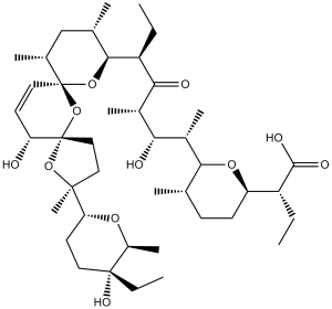
| Formula | C42H70O11 |
| CAS No. | 53003-10-4 |
| Storage | -20℃ for 3 years in powder form |
| -80℃ for 2 years in solvent | |
| Solubility (In vitro) | DMSO: <1 mg/mL |
| Water: <1 mg/mL | |
| Ethanol: <1 mg/mL | |
| SMILES | [H][C@]1([C@](C)(CC2)O[C@]32[C@H](O)C=C[C@]4(O[C@]([H])([C@@H](CC)C([C@@H](C)[C@@H](O)[C@H](C)[C@]5([H])O[C@]([C@@H](CC)C(O)=O)([H])CC[C@@H]5C)=O)[C@@H](C)C[C@H]4C)O3)CC [C@@](CC)(O)[C@H](C)O1 |
| Synonyms | Procoxacin; BioCox; Sacox; SalocinAHR-3096; AHR 3096; AHR3096; HSDB 7032; HSDB7032; HSDB-7032; K 364; Salinomycin; Coxistac. |
| In Vitro | In vitro activity: Salinomycin (Procoxacin) is an antibacterial and coccidiostat ionophore therapeutic agent. Salinomycin (Procoxacin) has been shown by Piyush Gupta to kill breast cancer stem cells at least 100 times more effectively than another popular anti-cancer compound (paclitaxel) in mice. The mechanism of action by which salinomycin (Procoxacin) kills cancer stem cells specifically remains unknown, but is thought to be due to its action as a potassium ionophore due to the detection of Nigericin in the same compound screen. Salinomycin has high toxicity and a narrow therapeutic window which may limit its clinical use. Kinase Assay: Salinomycin is a potent inhibitor of the Wnt signaling cascade. Incubation of the malignant lymphocytes with Salinomycin induces apoptosis within 48 h, with a mean IC50 of 230 nM. Salinomycin is also an antibiotic potassium ionophore, has been reported recently to act as a selective breast cancer stem cell inhibitor. Salinomycin is a novel and an effective anticancer drug, inhibits SW620 cells and Cisp-resistant SW620 cells with IC50 of 1.54±0.23 μM and 0.32±0.05 μM, respectively. Salinomycin is found to have the ability to kill both cancer stem cells (CSCs) and therapy-resistant cancer cells. After continuous Salinomycin treatment for 48 h, the apoptotic cells are observed under the microscope and counted randomly at least 100 cells in one field. The number of apoptotic cells which are stained by Hoechst33342 is significantly increased in Cisp-resistant SW620 cells (20.20±3.72) than that of SW620 cells (9.40±2.07) per 100 cells (p<0.05). After treatment with Salinomycin for 48 h, flow cytometric analysis is used to detect the cell apoptosis both in SW620 cells and Cisp-resistant SW620 cells. The cell apoptotic rate in Cisp-resistant SW620 cells (37.82±3.63%) is significantly higher than that of SW620 cells (16.78±2.56%) (p<0.05). Cell Assay: For cisplatin or Salinomycin IC50 analysis in SW620 cells or Cisp-resistant SW620 cells, cells (1×10⁴/well) are cultured in 96-well plates and treated with different chemotherapeutics (cisplatin, Salinomycin) in different concentrations for 48 h. Then 20 μL of cell counting kit-8 (CCK-8) is added into each of the 96-wells. After 4 h incubation at 37°C, the optical density (OD) values are detected at 450 nm using the scan reader. Cell growth inhibiting rates are described as cell inhibiting curves and the IC50 parameters (inhibiting concentration of 50% cells) are evaluated by Xlfit 5.2 software. For cell proliferation analysis, SW620 cells or Cisp-resistant SW620 cells (5×10³/well) are also seeded in 96-well plates in serum-containing medium and treated with cisplatin (5 μM, according to the calculated IC50 values of cisplatin in SW620 cells) for 0, 12, 24, 48, 72 and 96 h. Then 20 μL cell counting kit-8 is added into each of the 96-wells. After 4-h incubation at 37°C, the coloring reactions are also quantified at 450 nm. Several hepatocellular carcinoma (HCC) cell lines were treated with Sal. Results showed that Sal inhibited proliferation and decreased PCNA levels. Cell cycle analysis showed that Sal caused cell cycle arrest in different phases. Sal induced apoptosis as characterized by an increase in the Bax/Bcl-2 ratio. Compared to control, β-catenin expression was down-regulated by Sal treatment significantly. The Ca2+ concentration in HCC cells was examined by flow cytometry and it was found that higher Ca2+ concentrations were observed in Sal treatment groups. |
|---|---|
| In Vivo | The in vivo anti-tumor effect of Sal was verified using the hepatoma orthotopic tumor model and results showed that the liver tumor size in Sal-treated groups decreased. Immunohistochemistry and TUNEL staining also demonstrated that Sal could in vivo inhibit proliferation and induced apoptosis. |
| Animal model | Mice: Nude mice (nu/nu; 4-6 weeks of age) are used. HepG2 cells are suspended in 100 mL 1:1 serum-free DMEM and Matrigel. Mice are anesthetized with ketamine/xylazine and after surgically opening the abdomen, HepG2 cells are inoculated into the liver parenchyma and mice are monitored every 3 days for 35 days. Finally, 18 nude mice are divided into three groups that are intraperitoneally injected daily for 6 weeks: two Salinomycin-treated groups (4 mg/kg Salinomycin group, 8 mg/kg Salinomycin group) and the control group (saline water group) Rats: total of 10 male rats are used in the experiment. After a routine anesthesia, the abdomen is opened. After a resuspension of high glucose medium not containing serum DMEM, and matrigel, the bladder transitional cancer cell line T24 is inoculated in the parenchyma of bladder in rats, and then the abdomen is sutured. After operation, the rats are randomized into the experiment group and the control group with five in each group. After operation, the rats in the experiment group are immediately given intraperitoneal injection of Salinomycin with a dosage of 8 mg/kg, while the rats in the control group are given intraperitoneal injection of normal saline. A close observation is paid during the drug administration period. After 15 d, the rats are sacrificed by cervical dislocation, and the complete tumor tissues are stripped to observe the tumor growth and metastasis. |
| Formulation & Dosage | Mice: 4 and 8 mg/kg, i.p. inection; Rat: 8 mg/kg, i.p. inection |
| References | J Antibiot (Tokyo). 1974 Nov;27(11):814-21; Chem Biol Drug Des. 2012 Mar;79(3):235-8. |
 m.cnreagent.com