| CAS NO: | 103404-90-6 |
| 规格: | ≥98% |
| 包装 | 价格(元) |
| 5mg | 电议 |
| 10mg | 电议 |
| 25mg | 电议 |
| 50mg | 电议 |
| 100mg | 电议 |
| 250mg | 电议 |
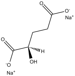
| Molecular Weight (MW) | 192.08 |
|---|---|
| Formula | C5H6Na2O5 |
| CAS No. | 103404-90-6 |
| Storage | -20℃ for 3 years in powder form |
| -80℃ for 2 years in solvent | |
| Solubility (In vitro) | DMSO: <1 mg/mL |
| Water: 38 mg/mL (197.83 mM) | |
| Ethanol: <1 mg/mL | |
| SMILES Code | O=C([O-])[C@H](O)CCC([O-])=O.[Na+].[Na+] |
| Synonyms | D-alpha-Hydroxyglutaric acid disodium salt; Disodium (R)-2-hydroxyglutarate; D-2-HGA; 2R-hydroxy-pentanedioic acid, disodium salt Chemical Name: 2R-hydroxy-pentanedioic acid, disodium salt |
| In Vitro | In vitro activity: Disodium (R)-2-Hydroxyglutarate (also known as D-2-HGA, or 2-HG) is a potent and competitive inhibitor of α-ketoglutarate (α-KG)-dependent dioxygenases with Ki value of 0.628 mM. In U-87MG cells, (R)-2-Hydroxyglutarate acts as weak antagonists of α-KG to inhibit α-KG-dependent histone demethylases and increases dimethylation on both H3K9 and H3K79. Besides, (R)-2-Hydroxyglutarate also inhibits ATP synthase and mTOR signaling, and therefore causes growth arrest and tumor cell killing. Inhibition of ATP synthase by 2-HG or α-KG in glioblastoma cells is sufficient for growth arrest and tumor cell killing under conditions of glucose limitation, e.g., when ketone bodies (instead of glucose) are supplied for energy. These findings inform therapeutic strategies and open avenues for investigating the roles of 2-HG and metabolites in biology and disease. Kinase Assay: To assay human JHDM1A/KDM2A demethylase activity toward H3K36me2, His tagged JHDM1A is first obtained by transforming pET28a-JHDM1A into Escherichia coli BL21 and protein expression is induced by addition of 1 mM IPTG at 30° C when cell density reaches 0.5 OD600 units. Cells are lysed by sonication and Ni-NTA agarose is used to purify His-JHDM1A fusion proteins. Histone demethylase assay is carried out by incubating 2 μg oligonucleosomes, 4 μg purified His-JHDM1A, and/or 10–50 mM L- or D-2-HG in histone demethylation buffer [50 mM HEPES (pH 8.0), 625 μM Fe(NH4)2(SO4)2, 0.1–0.5 mM α-KG, 2 mM ascorbate] at 37° C for 2–3 hr and the reactions are stopped by the addition of SDS loading buffer and subsequently analyzed by western blotting using anti-H3K36me2 antibody. To measure CeKDM7A demethylase activity toward H3K9me2 and H3K27me2, two synthetic dimethylated peptides H3K9me2 [ARTKQTARK (me2)STGGKA] and H3K27me2 [QLATKAARK (me2)SAPAS] are used as substrates. Demethylase assays are carried out in the presence of 10 μg enzyme, 1 μg peptide in 20 μl buffer 20 mM Tris-HCl (pH 7.5), 150 mM NaCl, 50 μM Fe(NH4)2(SO4)22, 100 μM α-KG, 2 mM Vc, 10 mM PMSF for 3 hr. The demethylation reaction mixture is desalted by passing through a C18 ZipTip. To examine the inhibitory effect of 2-HG, various concentrations of 2-HG are incubated with KDM7A briefly before adding other reaction mixtures. The samples are analyzed by a MALDI-TOF/TOF mass spectrometer. Cell Assay: Cells are seeded in 12-well plates and, after overnight incubation, treated with the indicated concentrations of each compound. After harvesting, cells are stained with acridine orange (AO) and 4′,6-diamidino-2-phenylindole (DAPI). Cell number and viability are measured based on AO and DAPI fluorescence as measured by NC3000 following the manufacturer's instructions. |
|---|---|
| In Vivo | |
| Animal model | |
| Formulation & Dosage | |
| References | Cancer Cell. 2011 Jan 18;19(1):17-30; Cell Metab. 2015 Sep 1;22(3):508-15. |
 m.cnreagent.com