| 规格: | 98% |
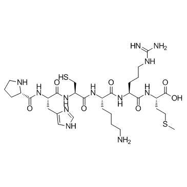
| 分子量: | 770.97 |
| 包装 | 价格(元) |
| 1mg | 电议 |
| 5mg | 电议 |
| 10mg | 电议 |
Background:
Antioxidant peptide A is a short peptide, which contains alternative aromatic or sulfur-containing amino acid. The side chains of Antioxidant peptide A are believed to contribute to strong radical scavenging activities of peptides in the cancer cell.
The effects of 10-100 μM of Antioxidant peptide A (Pep-A) concentrations are studied on the superoxide dismutase (SOD) enzyme activity. The enzyme activity decreases by 0.5 and 0.7-folds at 10 and 50 µM Antioxidant peptide A concentrations, respectively, and increases by 1.79-folds at 100 µM Antioxidant peptide A treatment, indicating that this concentration can be ideal for the treatment on Y79 a, RB cells. Furthermore, the Antioxidant peptide A can be involved in decreasing the ROS by increasing the antioxidant enzyme activity. A similar increase in the antioxidative enzyme levels in the presence of Hoki skin antioxidative peptide in hepatocarcinoma cells is attributed to the peptide's role in maintaining the redox balance in the cellular environment. Cell viability analysis results show that the Antioxidant peptide A shows no toxicity to cancerous (Y79) cells and non-cancerous cells even after 48 h of treatment. The Y79 RB cell viability ranges between 115 and 157 % and 111-126 % after 24 and 48 h of exposures with Antioxidant peptide A, respectively. The cancer cell death from the treatment of 10-100 µM GNPs concentration is studied[1].
[1]. Kalmodia S, et al. Bio-conjugation of antioxidant peptide on surface-modified gold nanoparticles: a novel approach to enhance the radical scavenging property in cancer cell. Cancer Nanotechnol. 2016;7:1.
Protocol:
Kinase experiment: | Y79 cells are seeded at 2×10⁵ cells/well in 12-well plates with 1000 μL of culture media and incubated at 37°C overnight. The cells are then exposed to varying dosages of Antioxidant peptide A (Pep-A) and Pep-B (10, 50, and 100 μM) in fresh medium and then incubated for 6 h. At the end of the incubation, the cells are collected and washed with ice-cold PBS twice and lysed with cell lysis buffer [0.1 M Tris/HCl, pH 7.4 containing 0.5 % Triton X-100, 5 mM β-Mercaptoethanol, 0.1 mg/mL PMSF]. Cell lysate is centrifuged at 14000×g for 5 min at 4°C. The supernatant contains a combined SOD activity from cytosol and mitochondria. The SOD activity is measured using the superoxide dismutase (SOD) activity assay kit. In brief, the cell lysate, buffer, enzyme, and the WST reagent are diluted, and the solution is added as per the protocol and incubated at 37°C for 20 min and read at 450 nm on a microplate reader. SOD activity is then calculated as a percentage of the inhibition activity of xanthine oxidase[1]. |
Cell experiment: | Y79 retinoblastoma and MIOM1 cells are seeded at 5×10³ cells/well in 96-well plates and incubated at 37°C overnight. The cells are then treated with varying concentrations (10, 30, 60, and 100 μM) of Antioxidant peptide A (Pep-A) and Pep-B in a fresh medium and incubated for specific time periods (24 and 48 h). At the end of the incubation, 10 μL of MTT (5 mg/mL) is added to the cells with fresh medium (100 μL) and incubated at 37°C until formazan crystals are formed. The formazan crystals are dissolved in 100 μL of DMSO, and the reading is taken at 570 nm. Cell viability is calculated[1]. |
参考文献: [1]. Kalmodia S, et al. Bio-conjugation of antioxidant peptide on surface-modified gold nanoparticles: a novel approach to enhance the radical scavenging property in cancer cell. Cancer Nanotechnol. 2016;7:1. | |
 m.cnreagent.com