| 包装 | 价格(元) |
| 5mg | 电议 |
| 25mg | 电议 |
| 100mg | 电议 |
Kinase experiment: | A PI3K Alphascreen assay is used to measure the activity of a panel of four phosphoinositide 3-kinases: PI3Kα, PI3Kβ, PI3Kγ, and PI3Kδ. Enzyme reaction buffer is prepared using sterile water and 50 mM Tris-HCl, pH 7, 14 mM MgCl2, 2 mM sodium cholate, and 100 mM NaCl. 2 mM DTT is added fresh on the day of the experiment. The Alphascreen buffer is made using sterile water and 10 mM Tris-HCl, pH 7.5, 150 mM NaCl, 0.10% Tween 20, and 30 mM EDTA. Then 1 mM DTT is added fresh on the day of the experiment. Compound source plates used for this assay are 384-well Greiner clear polypropylene plates containing test compounds at 5 mM and diluted 1:2 over 22 concentrations. Columns 23 and 24 contained only DMSO, as these wells comprised the positive and negative controls, respectively. Source plates are replicated by transferring 0.5 μL per well into 384-well Optiplates. Each PI3K isoform is diluted in enzyme reaction buffer to 2× working stocks. PI3Kα is diluted to 1.6 nM, PI3Kβ is diluted to 0.8 nM, PI3Kγ is diluted to 15 nM, and PI3Kδ is diluted to 1.6 nM. PI(4,5)P2 is diluted to 10 μM, and ATP is diluted to 20 μM. This 2× stock is used in the assays for PI3Kα and PI3Kβ. For assay of PI3Kγ and PI3Kδ, PI(4,5)P2 is diluted to 10 μM and ATP is diluted to 8 μM to prepare a similar 2× working stock. Alphascreen reaction solutions are made using beads from the anti-GST Alphascreen kit. Two 4× working stocks of the Alphascreen reagents are made in Alphascreen reaction buffer. In one stock, biotinylated-IP4 is diluted to 40 nM and streptavadin-donor beads are diluted to 80 μg/mL. In the second stock, PIP3-binding protein is diluted to 40 nM and anti-GST-acceptor beads are diluted to 80 μg/mL. The plates are incubated at room temperature for 30 min. Still in the dark, 10 μL/well of the acceptor bead solution is added to columns 1-24 of the plates. The plates are then incubated in the dark for 1.5 h. The plates are read on an Envision multimode plate reader using a 680 nm excitation filter and a 520-620 nm emission filter[1]. |
Cell experiment: | B cells are purified from human peripheral blood mononuclear cells (PBMCs) by negative selection. Approximately 3×10⁴ purified B cells per well are seeded into a 96-well plate. Compounds (e.g., AMG319) are dissolved in DMSO at a concentration of 10 mM, and a 10-point, 3-fold serial dilution of the compound is carried out in DMSO. Then 0.5 μL of compound is added to each well in duplicates so that the final DMSO concentration is 0.25% and the highest compound concentration is 10 μM. After preincubating for 30 min, B cells are treated with 2 μg/mL of anti-human IgM antibody plus 300 ng/mL human CD40L or 5 ng/mL human IL-4 plus 200 ng/mL of CD40L as a counterscreen to evaluate the off-target effects. The plates are incubated at 37℃ and 5% CO2 for 72 h, then pulsed with 0.5 μCi per well 3H thymidine for 18 h, and B cells are collected to count the incorporation of 3H thymidine[1]. |
Animal experiment: | Mice[1] IgM membrane only homozygous transgenic mice (6- to 12-week-old female) are orally dosed with AMG319 or vehicle control (n=5 per group). At 15 min after treatment, mice are tail iv injected with 50 μg of Endotoxin-free FITC-labeled μ chain specific anti-IgM or PBS only control. Blood and spleen tissue are collected after 30 min of stimulation for drug concentration and B cell pAKT analysis via flow cytometry. Briefly, blood and dispersed splenocytes are fixed with BD Phosflow lyse/fix buffer, pelleted, and permeabilized with cold 90% MeOH. Cells are then stained with pAKT and Alexa-647 secondary and B220-Pacific Blue for FACS analysis. Stimulated B220+/anti-IgM FITC+ B cells are analyzed for pAKT levels with B220+/FITC-B cells from anti-IgM untreated mice serving as a control. Mice are maintained and experiments are performed. Rats[1] Female Lewis rats (N=8/dose group) are dosed po with AMG319 or vehicle (2% HPMC, 1% Pluronic F68, 10% Captisol, pH 2.0) once a day for 10 days at various doses. Two hours after the first dosing, 200 μL of PBS containing 60 μg of KLH is administered to each rat intravenously. Ten days after the KLH priming, rats are euthanized and blood is taken by cardiac puncture for the measurement of KLH specific IgG and IgM by ELISA. |
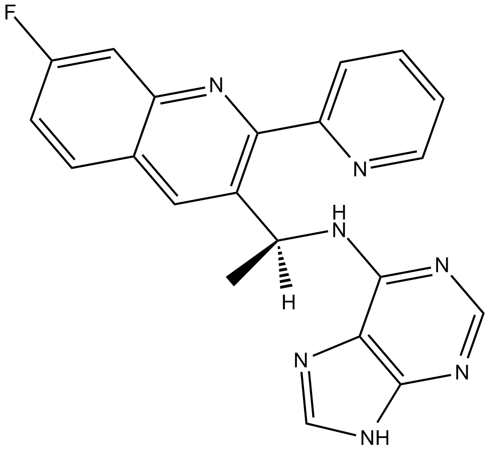
| 产品描述 | AMG 319, a highly selective inhibitor of phosphoinositide 3-kinase p110δ isoform (PI3Kδ) [1], with an IC50 value less than 10 nM [2]. PI3Kδ plays an essential role in B-cell receptor (BCR) signaling. PI3Kδ is expressed in lymphoid malignancies, including chronic lymphocytic leukemia (CLL) and non-Hodgkin lymphoma (NHL) [1]. C-Akt, a serine-threonine kinase is one target of PI39K. C-Akt is the prototypical member of a mammalian Akt isoform family. The regulation to Akt may be phosphorylation or direct binding the Akt pleckstrin homology domain with PI39K lipid products. PI39K-independent Akt stimuli had been identified [3]. AMG 319 inhibited basal AKT phosphorylation and proliferation in lymphoid tumor cells [1]. 28 patients received AMG 319. In a CLL patient after 1 dose of AMG 319, grade 3 hemolytic anemia at 25 mg was produced. All CLL samples with an inducible signal (60%) showed coverage of BCR-induced pAKT (ex-vivo IgD stimulated) dose-dependently; at 400 mg, near complete inhibition was seen for 24 hours. Baseline % of T-regulatory cells was elevated in CLL patients (14.4% ± 7.6%). But during treatment (14/19 patients), the elevated T regulatory cells tended to normalize. This suggested that the drug might produce immune restoration. By physical exam, all 20 evaluable patients showed greater than 50% lymph node (LN) reduction, 15 (75%) patients showed greater than 90% LN reduction. This response was present in all cytogenetic subtypes [1]. References: |
 m.cnreagent.com