| 包装 | 价格(元) |
| 1mg | 电议 |
| 5mg | 电议 |
| 10mg | 电议 |
Cell lines | HeLa, Vero, L, HEp2, and MDBK cells, SC-1 cells, Murine CT26 colorectal carcinoma cells |
Preparation method | The solubility of this compound in DMSO is > 10 mM. General tips for obtaining a higher concentration: Please warm the tube at 37 ℃ for 10 minutes and/or shake it in the ultrasonic bath for a while. Stock solution can be stored below -20℃ for several months. |
Reacting condition | 0.2–0.5 μg/ml |
Applications | In HeLa, Vero, L, HEp2, and MDBK cells, cytochalasin D (0.2–0.5 μg/ml) induced sustained contraction (contracture), loss of microvilli, expression of endoplasmic contents (zeiosis), nuclear protrusion, and extension of cytoplasmic processes. Cells in G1 were most sensitive to CD; responsiveness decreased progressively during early S and is least in mid S through G2. CD inhibited transport of [14C]deoxyglucose in HeLa. In SC-1 cells, Cytochalasin D treatment severely disrupted network organization, increased the number of actin filament ends, and led to the formation of filamentous aggregates or foci composed mainly of actin filaments. Cytochalasin D (0.24~15 μg/mL, 16 h) inhibited CT26 tumor cell proliferation in time and dose dependent manner and induced significant CT26 cell apoptosis. |
Animal models | Murine CT26 tumor model, porcine coronary model |
Dosage form | Intravenous injection, 50 mg/kg, every 3 days for 21 days |
Application | Cytochalasin D (i.v., 50 mg/kg) in vivo treatment significantly inhibited tumor growth and prolonged the survival times in CT26 tumor-bearing mice. In porcine coronary model, Cytochalasin D (2 μg) resulted in less late lumen loss in low-dose. High-dose Cytochalasin D (20 μg) inhibited both late lumen loss and intimal area. |
Other notes | Please test the solubility of all compounds indoor, and the actual solubility may slightly differ with the theoretical value. This is caused by an experimental system error and it is normal. |
| 文献引用 | |
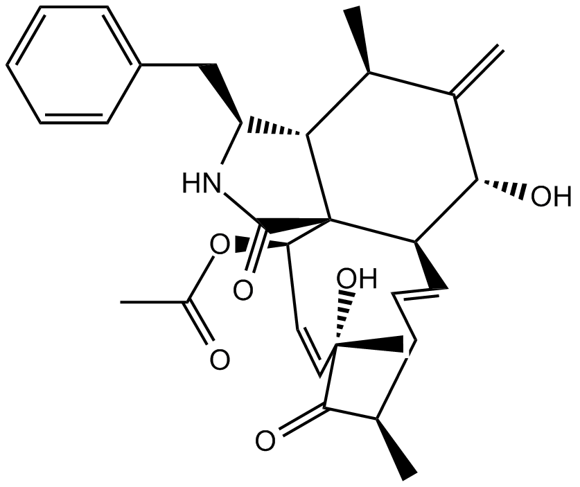
| 产品描述 | The cytochalasins are cell-permeable fungal metabolites that inhibit actin polymerization.[1],[2],[3],[4] This interferes with such diverse processes as cell movement, growth, phagocytosis, degranulation, and secretion.[5],[6],[7],[8] Cytochalasin D is a cell-permeable inhibitor that binds actin filaments, but not actin monomers, to inhibit polymerization at concentrations as low as 0.2 μM.2 In this way, it prevents the migration of tumor cells.[9] Reference: |
 m.cnreagent.com