| CAS NO: | 5142-23-4 |
| 规格: | ≥98% |
| 包装 | 价格(元) |
| 500mg | 电议 |
| 1g | 电议 |
| 2g | 电议 |
| 5g | 电议 |
Molecular Weight (MW) | 149.15 |
Formula | C6H7N5 |
CAS No. | 5142-23-4 |
Storage | -20℃ for 3 years in powder form |
-80℃ for 2 years in solvent | |
Solubility (In vitro) | DMSO: 3 mg/mL warming (20.11 mM) |
Water: 20 mg/mL warming (134.09 mM) | |
Ethanol: 4 mg/mL (26.81 mM) | |
Other info | Synonym: 3-Methyladenine, NSC 66389; NSC-66389; NSC66389. Chemical Name: 3-methyl-3H-purin-6-amine InChi Key: FSASIHFSFGAIJM-UHFFFAOYSA-N InChi Code: InChI=1S/C6H7N5/c1-11-3-10-5(7)4-6(11)9-2-8-4/h2-3H,7H2,1H3 SMILES Code: NC(N=CN1C)=C2C1=NC=N2 |
Chemical Name | 3-methylpurin-6-amine; 3-MA; 3MA; 3MA |
In Vitro | Kinase Assay: Protein degradation assay; HeLa cells are radiolabeled for 24 hours with 0.05 mCi/mL l-[U- 14C]valine. At the end of the labeling period, cells are rinsed three times with PBS. Cells are incubated for the designated times in either full medium or EBSS with or without the presence of 10 mM 3-Methyladenine.
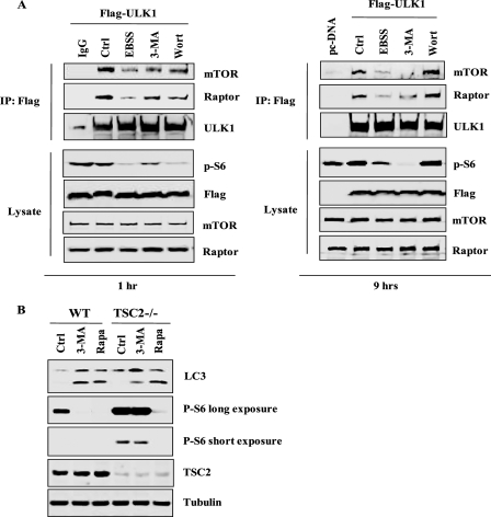
Cell Assay: Cell (such as HeLa cell) viability is determined by a trypan blue exclusion assay. Briefly, after treated with 3-Methyladenine, both adherent and floating cells are collected and suspended in phosphate buffered saline (PBS, pH 7.4) at a final density of 1-2 × 10⁶/mL. An equal volume of 0.4% trypan blue solution (w/v, in PBS) is added to the cell suspension and mixed thoroughly. After incubation at room temperature for 3 min, cell counting is performed using a hemacytometer. The slight preference for Vps34 prevention by 3-Methyladenine probably arises from a hydrophobic ring specific to Vps34, which encircles the 3-methyl group of 3-Methyladenine. 3-Methyladenine has been reported to cause cancer cell death under both normal and starvation conditions. 3-Methyladenine could also suppress cell migration and invasion independently of its ability to inhibit autophagy, implying that 3-Methyladenine possesses functions other than autophagy suppression. 3-Methyladenine elicits caspase-dependent cell death that is independent of autophagy inhibition. Treatment with 5 mM 3-Methyladenine reduces the percentage of glucose-starved HeLa cells displaying GFP-LC3 puncta to 23%. The levels of LC3-I are increasing and the levels of LC3-II are decreasing between 12 and 48 hours in cells that are treated with 3-Methyladenine. Conversion of LC3-I to LC3-II is suppressed by 3-Methyladenine. Treatment of HeLa cells with 3-Methyladenine at 2.5 mM or 5 mM for one day does not affect cell viability, whereas treatment with 10 mM 3-Methyladenine for one day causes a 25.0% decrease in cell viability. Treatment of cells with 2.5, 5 or 10 mM 3-Methyladenine for two days causes 11.5%, 38.0% and 79.4% decrease in viability, respectively. 3-Methyladenine decreases cell viability in a time- and dose-dependent manner. 3-Methyladenine significantly shortens the duration of nocodazole-induced-prometaphase arrest. Suppression of autophagy by 3-Methyladenine inhibits SU11274-induced cell death. Prolonged treatment with 3-Methyladenine (up to 9 hours) induces significant LC3 I to II conversion in wild type MEFs. Prolonged treatment with 3-Methyladenine, but not wortmannin, markedly increases GFP-LC3 punctuation/aggregation. 3-Methyladenine-induced LC3 conversion and free GFP liberation are ATG7-dependent. 3-Methyladenine treatment leads to evident increase of p62 protein level. 3-Methyladenine increases the p62 level even in Atg5–/– MEFs as well as in cells with DOX-mediated deletion of ATG5. 3-Methyladenine inhibits class I and class III PI3K in different temporal patterns. 3-Methyladenine-induced LC3 I to LC3 II conversion is dramatically compromised in Tsc2–/– cells compared with wild type cells.3-Methyladenine disrupts the anti-autophagic function of mTOR complex 1. |
In Vivo | 3-Methyladenine blocks autophagy through its effect on class III phosphatidylinositol 3-kinase (PI3K). 3-Methyladenine treatment does not alter the degree of hemorrhage compared with the subarachnoid hemorrhage (SAH) group. 3-Methyladenine pretreatment significantly aggravates neurological symptoms when compared with the SAH + vehicle group. Autophagy is decreased when 3-Methyladenine treatment is applied. Conversely, cleaved caspase-3 is markedly up-regulated in the SAH + 3-Methyladenine group. In line with the up-regulation of cleaved caspase-3 expression, the number of TUNEL-positive cells in the right cortex is significantly increased in the SAH + 3-Methyladenine group compared with the SAH + vehicle group |
Animal model | Adult male Sprague–Dawley rats weighing 300-350 g |
Formulation & Dosage | Dissolved in saline.;400 nM; Intracerebral ventricular |
References | [1] Miller S, et al. Autophagy. 2010, 6(6), 805-807.; [2] Jing CH, et al. Neuroscience. 2012. |
Prolonged treatment with 3-MA in full medium leads to accumulation of autophagic markers. J Biol Chem. 2010 Apr 2;285(14):10850-61. |
3-MA increases autophagic flux. J Biol Chem.2010 Apr 2;285(14):10850-61. |
3-MA does not affect lysosomal functions. J Biol Chem. 2010 Apr 2;285(14):10850-61. |
3-MA inhibits class I and class III PI3K in different temporal patterns. J Biol Chem. 2010 Apr 2;285(14):10850-61. |
3-MA disrupts the function of mTOR complex I. J Biol Chem. 2010 Apr 2;285(14):10850-61. |
|
 m.cnreagent.com