| 包装 | 价格(元) |
| 10mM (in 1mL DMSO) | 电议 |
| 20mg | 电议 |
Cell experiment: | HBZY-1 cells are plated into 96-well plates and pretreated with various concentrations(1, 5, 10, 25, 50, 100 μM) of naringin for 2 h. Then cells are treated with 30 mM glucose for 24 h. The control group is added sterile normal saline in the same volume. After treatment, all the wells are incubated with 20 μL of 5 mg/ml MTT for 4 h at 37℃. Subsequently, 100 μL of DMSO are used to dissolve the formed formazan crystals after removal of the supernatant. The result is recorded at 490 nm on a microplate reader[1]. |
Animal experiment: | Rats: The rats are randomly divided into six groups: control, naringin (80 mg/kg), STZ, STZ+naringin (20 mg/kg), STZ+naringin (40 mg/kg), STZ+naringin(80 mg/kg). The rats in the STZ and STZ+naringin groups are intraperitoneally injected with STZ (65 mg/kg). The control and naringin groups are intraperitoneally injected with 0.1 M citrate buffer of same volume. After injection of STZ for 3 and 5 days, blood glucose levels are measured by tail vein puncture blood sampling[1]. Mice: Sixty 4-week-old male mice are randomized into four groups and fed for 20 weeks with either control diet or high-fat diet chow. Mice are dosed with 100 mg/kg of naringin daily. Mice body weight and food intake are weekly measured. Following behavioral assessment, animals are deeply anesthetized with isoflurane and sacrificed by decapitation after fasting for at least 5 h. Their plasma is collected for further analysis[4]. |
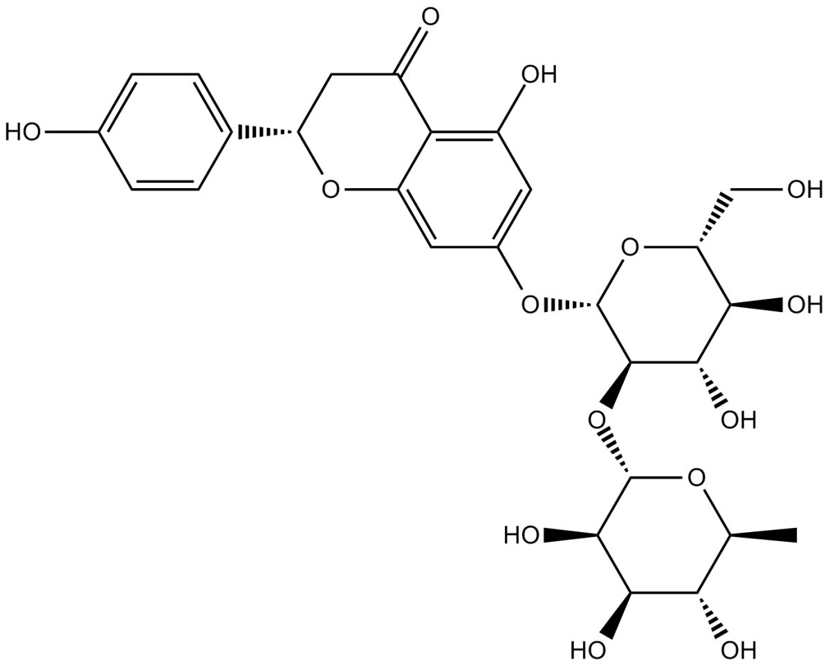
| 产品描述 | Naringin is a major flavanone glycoside obtained from tomatoes, grapefruits, and many other citrus fruits. Naringin exhibits biological properties such as antioxidant, anti-inflammatory, and antiapoptotic activities. Naringin suppresses NF-κ B signaling pathway activation. Naringenin inhibits high glucose-induced proliferation, inflammatory reaction and oxidative stress injury in HBZY-1 cells[1]. Naringin inhibits AGS cancer cell proliferation in a dose- and time-dependent manner. Phosphorylation of PI3K and its activated downstream targets p-Akt and p-mTOR are significantly decreased at 2 mM in Naringin-treated AGS cells. Naringin induces autophagic cell death in AGS cells. Naringin activated the autophagy related protein in AGS cells[2]. Naringin protects PC12 cells from 3-NP neurotoxicity. The lactate dehydrogenase release is decreased upon naringin treatment in 3-NP-induced PC12 cells. Naringin treatment enhances the antioxidant defense by increasing the activities of enzymatic antioxidants and the level of reduced glutathione[3]. Treatment with naringin significantly alleviates renal injury in diabetic rats and increases diabetic rats body weight significantly. Administration of naringin effectively alleviates the collagen deposition and renal interstitial fibrosis in diabetic rats. Treatment with naringin could result in decreased levels of ROS and MDA and increased activities of SOD and GSH-Px[1]. Oral administration of naringin significantly improves the learning and memory abilities. Naringin significantly enhances insulin signaling pathway[3]. References: |
 m.cnreagent.com