| 包装 | 价格(元) |
| 10mM (in 1mL DMSO) | 电议 |
| 10mg | 电议 |
Animal experiment: | Rats: The freshly isolated hepatocytes are preincubated for 2 h at a density of 1× 10⁶ cells/mL in a mixture of William’s E medium supplement with 10% FBS. Isolated rat hepatocytes are incubated in William’s E medium with or without (used as a control) GCDCA (50, 100 and 300 μM), or TG (1, 2 and 5 μM) for 1-24 h[3]. |
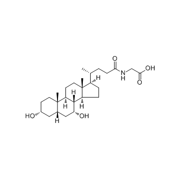
| 产品描述 | Glycochenodeoxycholic acid is a bile salt formed in the liver from chenodeoxycholate and glycine; used to induce hepatocyte apoptosis in research. Chenodeoxycholate is toxic to hepatocytes, and accumulation of chenodeoxycholate in the liver during cholestasis may potentiate hepatocellular injury. At a concentration of 250μM, glycochenodeoxycholate is more toxic than either chenodeoxycholate or taurochenodeoxycholate. Glycochenodeoxycholate cytotoxicity may result from ATP depletion followed by a subsequent rise in Ca2+. The rise in Ca2+ leads to an increase in calcium-dependent degradative proteolysis and, ultimately, cell death[1]. 4 h exposure of 50 μM GCDC induces apoptosis in 42% of hepatocytes. Intracellular PKC activity decreased to 44% of controls 2 h after exposure of hepatocytes to GCDC. GCDC-induced apoptosis is associated with decreases in total cellular PKC activity, which appear to be dependent on intracellular calpain-like protease activity[2]. GCDCA induces ER-related calcium release within about ten seconds. Significant increases in activities of calpain and caspase-12 are observed after 15 h of GCDCA treatment. Bip and Chop mRNA expressions are increased with the treated GCDCA dose and incubation time. Cytochrome c release from mitochondria peaks in about 2 h of incubation[3]. [1]. Spivey JR, et al. Glycochenodeoxycholate-induced lethal hepatocellular injury in rat hepatocytes. Role of ATP depletion and cytosolic free calcium. J Clin Invest. 1993 Jul;92(1):17-24. [2]. Gonzalez B, et al. Glycochenodeoxycholic acid (GCDC) induced hepatocyte apoptosis is associated with early modulation of intracellular PKC activity. Mol Cell Biochem. 2000 Apr;207(1-2):19-27. [3]. Tsuchiya S, et al. Involvement of endoplasmic reticulum in glycochenodeoxycholic acid-induced apoptosis in rathepatocytes. Toxicol Lett. 2006 Oct 10;166(2):140-9. |
 m.cnreagent.com